Plasmacytoma | Cases
Published on Aug 9, 2021
Retrospective case study
66 Male
- 1 year history of right thigh pain
- Attended physiotherapy. Given exercises for tight thigh muscle. No improvement.
- Consulted GP 10-12 months after physiotherapy as he developed problems with sit to stand.
- GP requested X-ray pelvis. X-ray report as follows. GP referral for x-ray mentions – “Injury right leg two years ago (jumped down and rounded on extended legs). Pain right side since then. Now limping.”
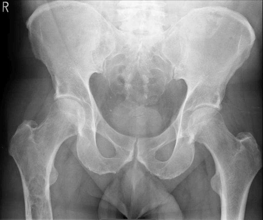
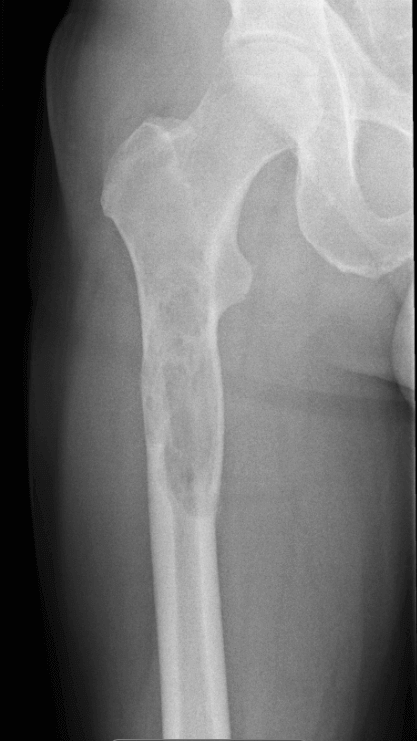
X-ray Pelvis
Well-defined lucent radiolucent lesion in the right upper femur, occupying most of the width of the femoral shaft, commencing about 12 cm below the level of the hip joint. Narrow zone of transition and endosteal scalloping. The inferior aspect of this lesion is not included on the film. We will recall the patient for further views of the right femur show the whole lesion and a second report/addendum will be issued. ( This patient will very probable require orthopaedic referral. Bone scintigraphy and/or MRI evaluation will probably be necessary.)
Further x-ray femur is then requested which gets done a week later.
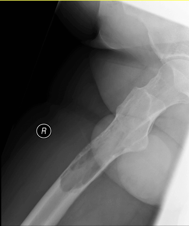
XR Femur (Thigh) R
There is an expansile 12cm lytic lesion in the diaphysis of the right femur with a scalloped border and narrow zone of transition. There is a ground glass appearance to the majority of the matrix. There is some focal cortical destruction and periosteal reaction. No other bone lesion.
A week later patient gets an MRI done for his thigh
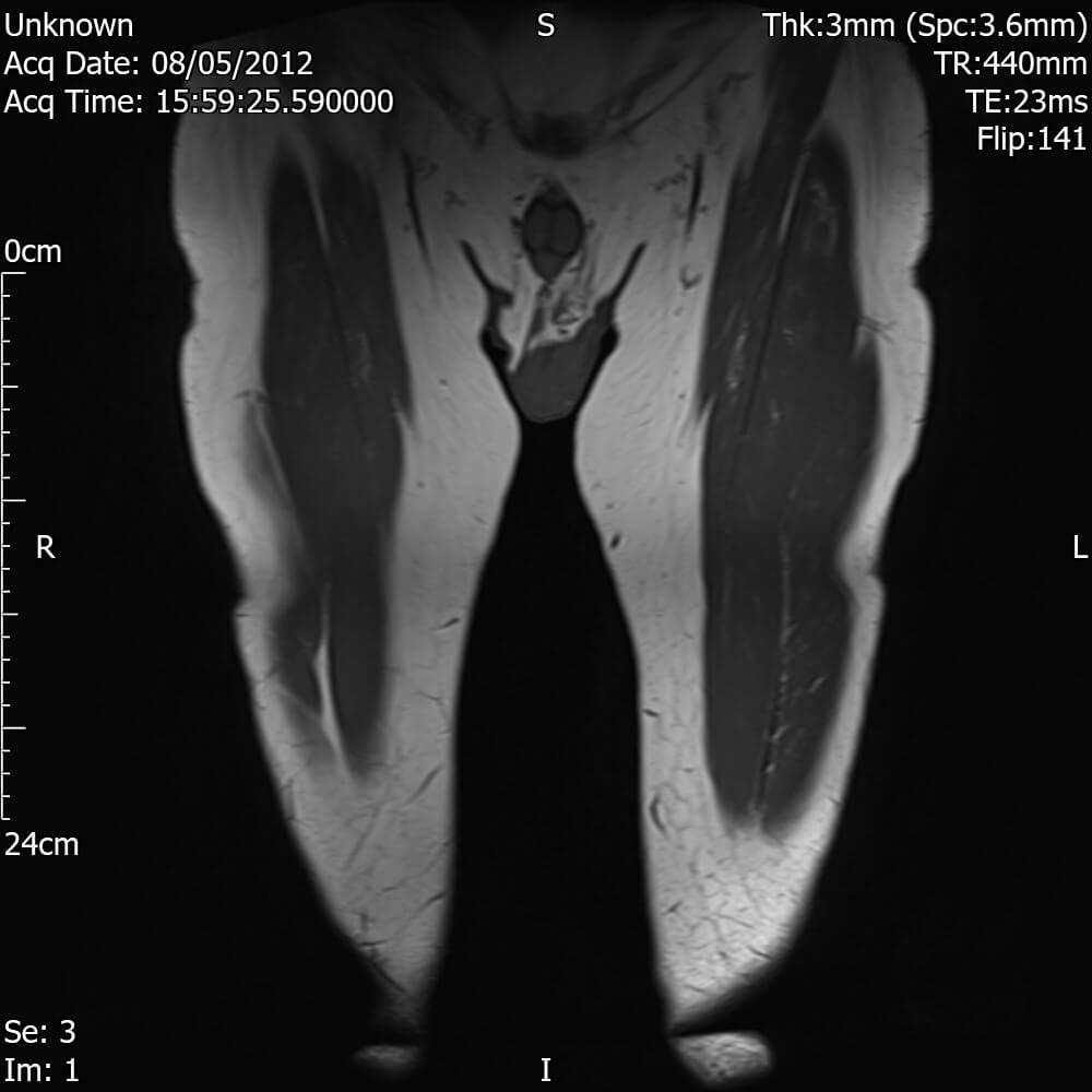
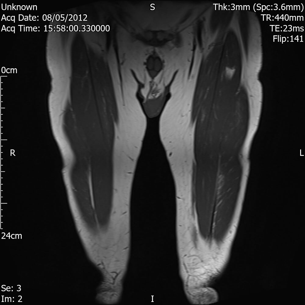
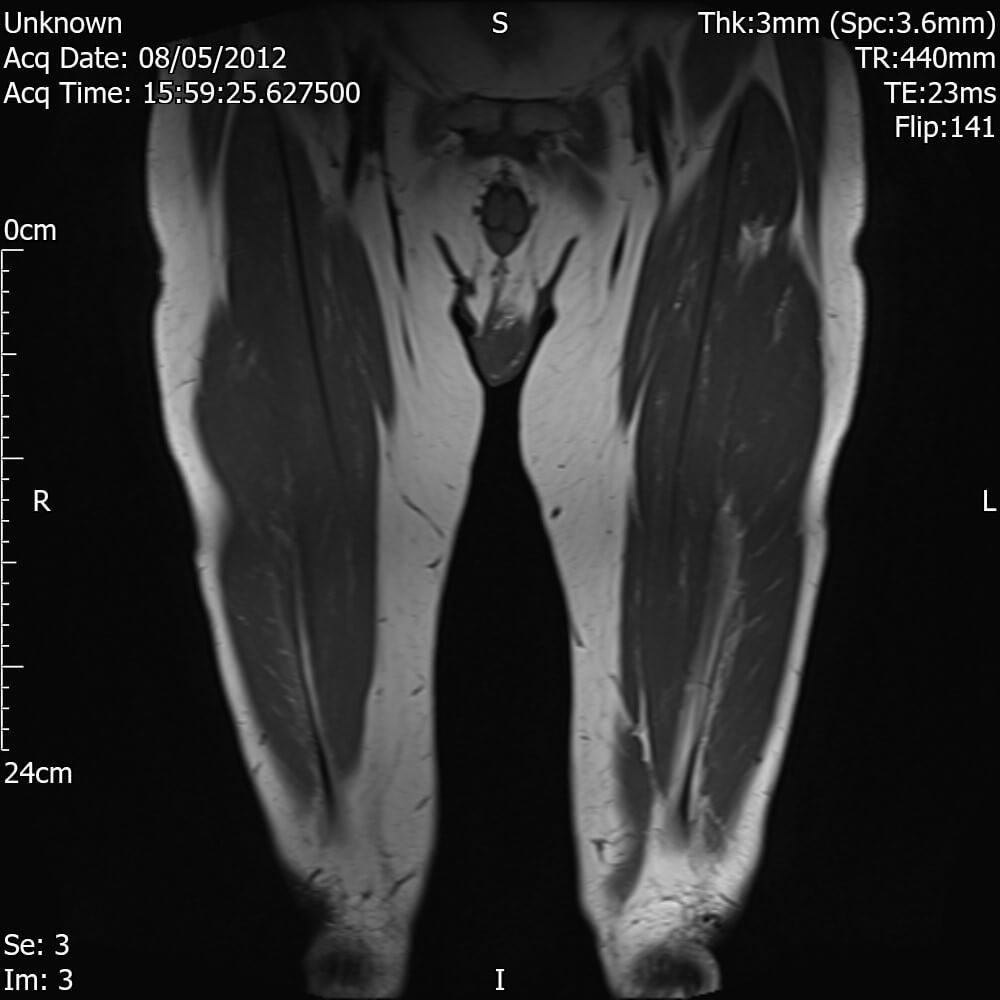
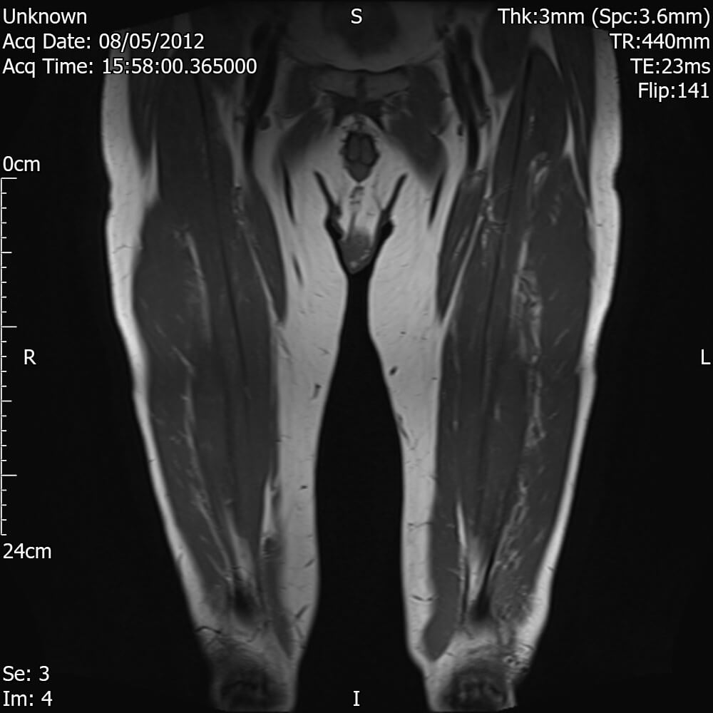
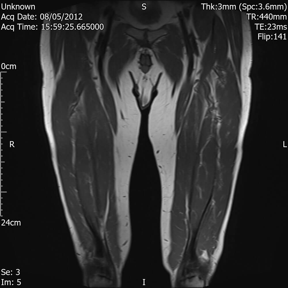
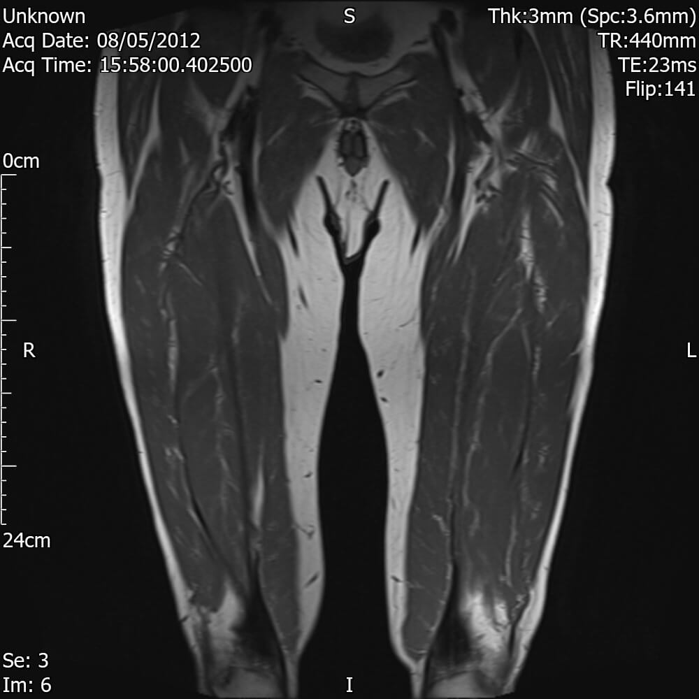
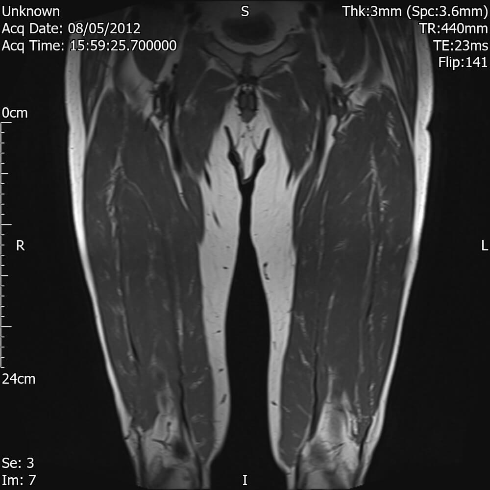
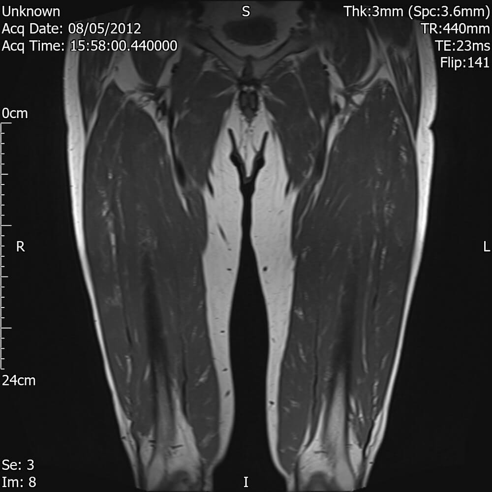
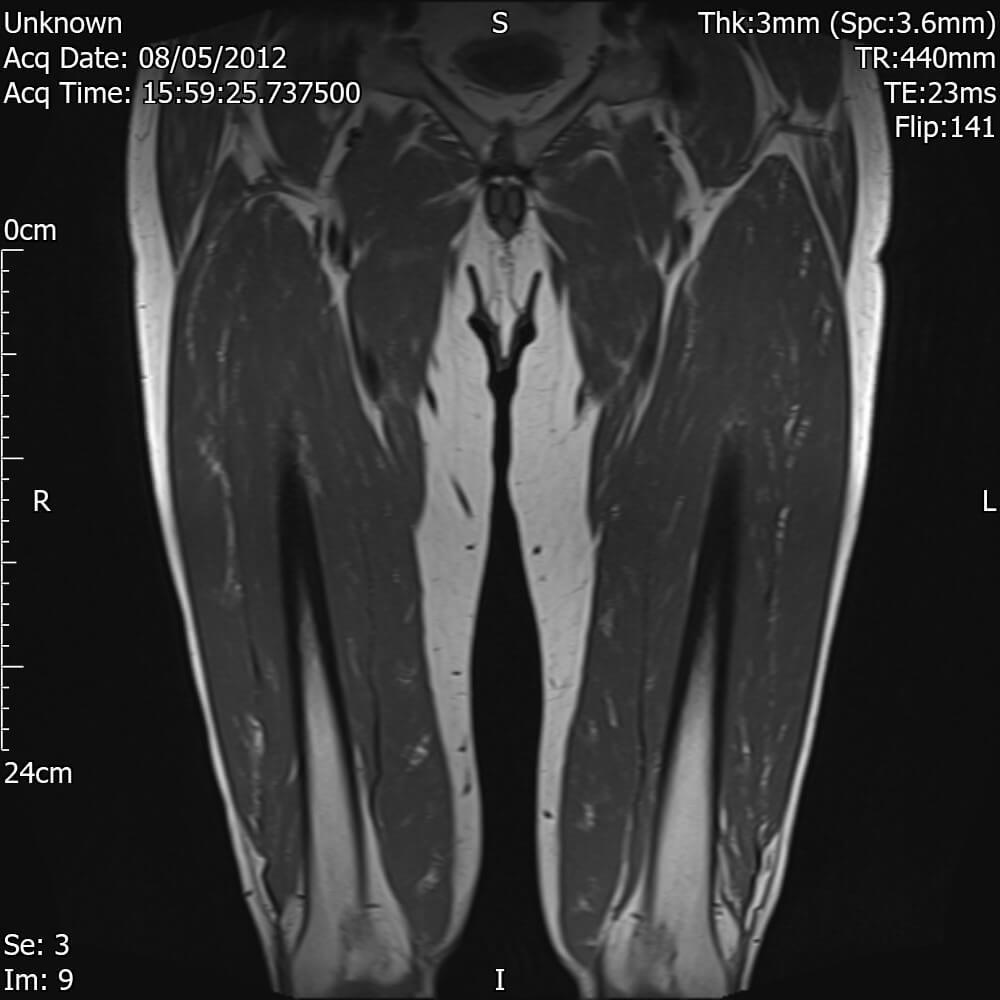
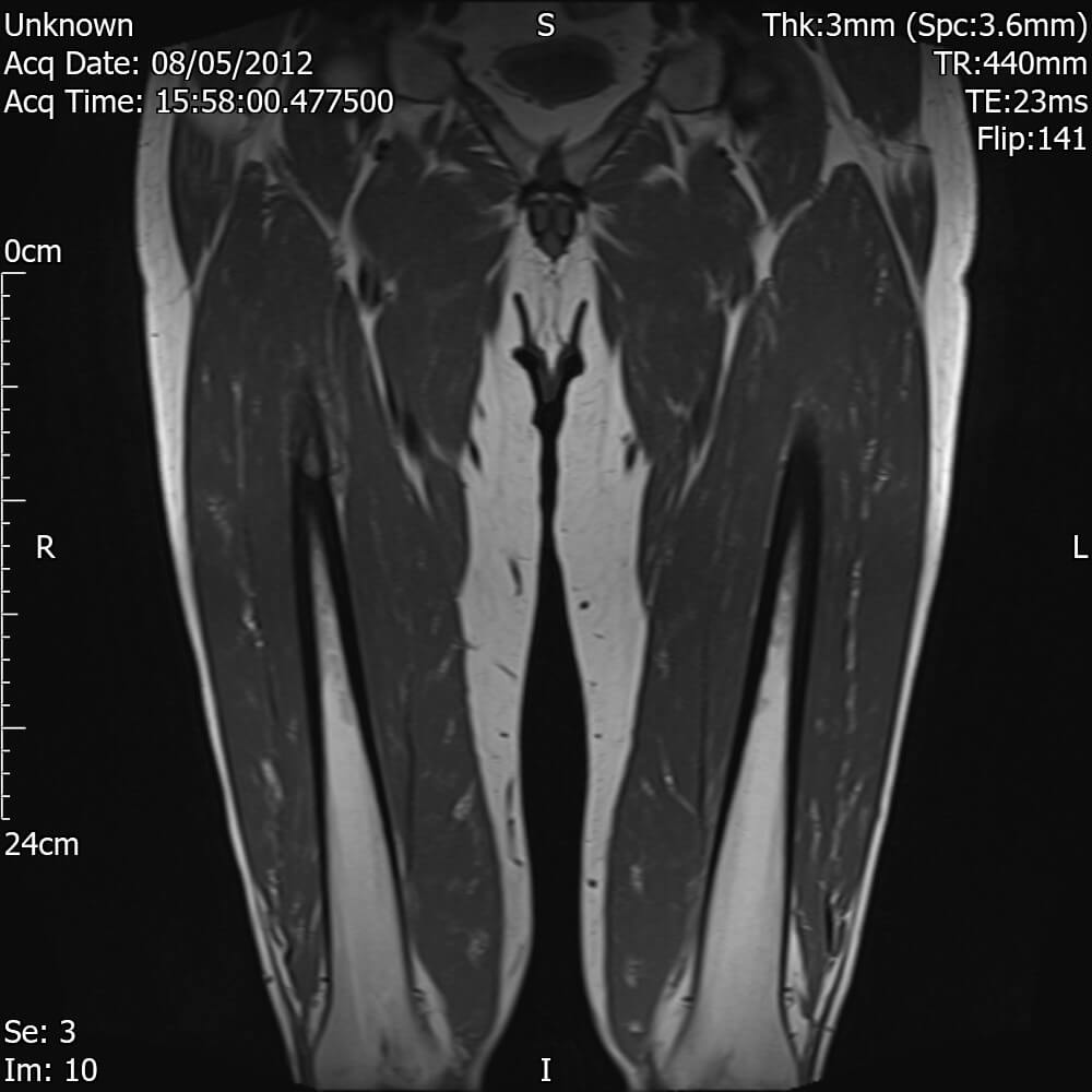
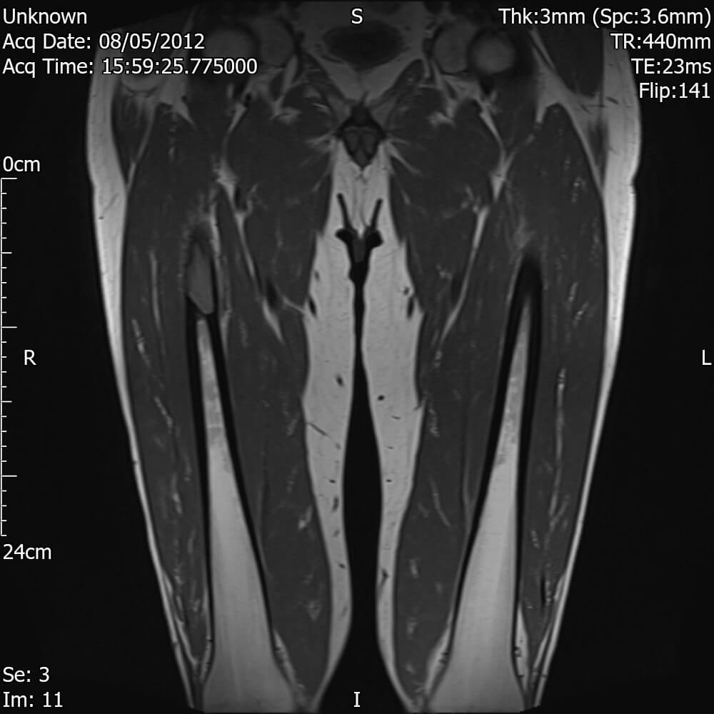
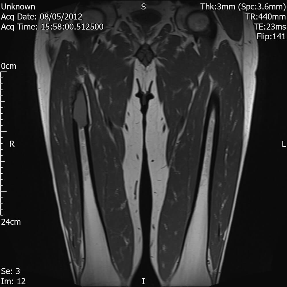
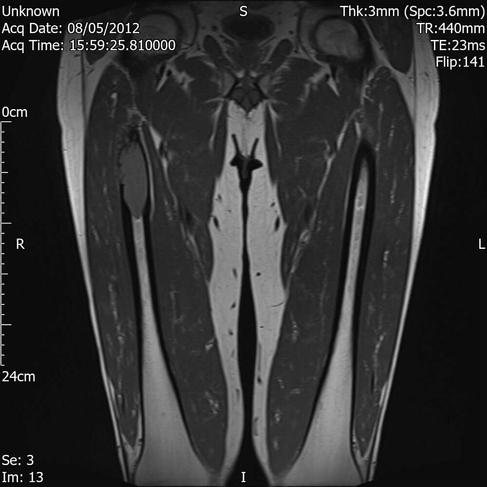
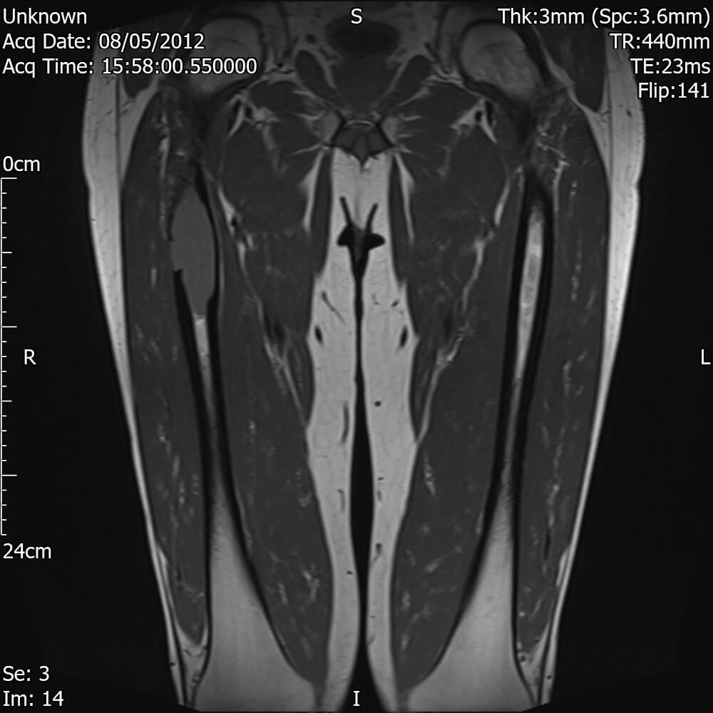
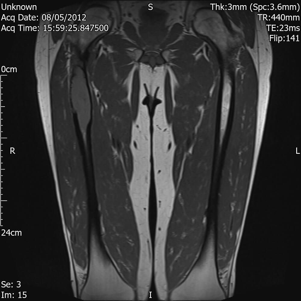
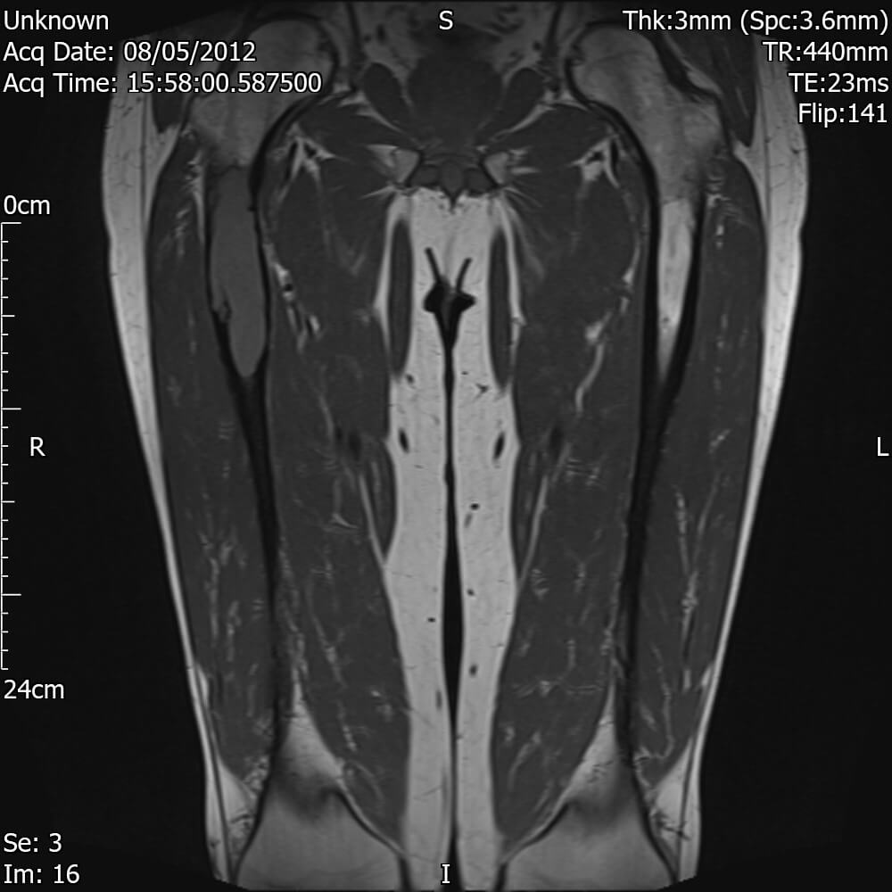
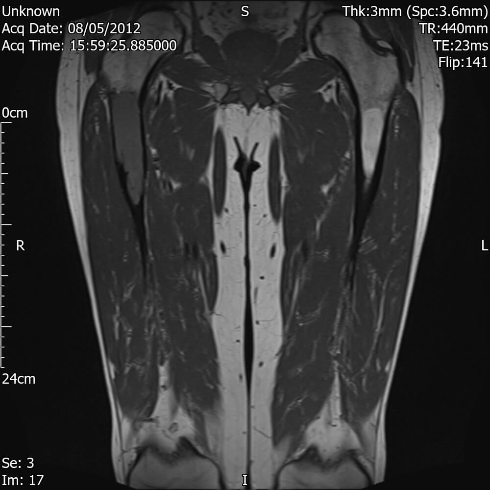
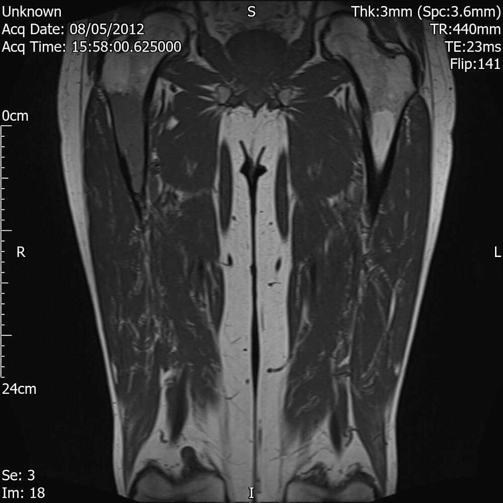
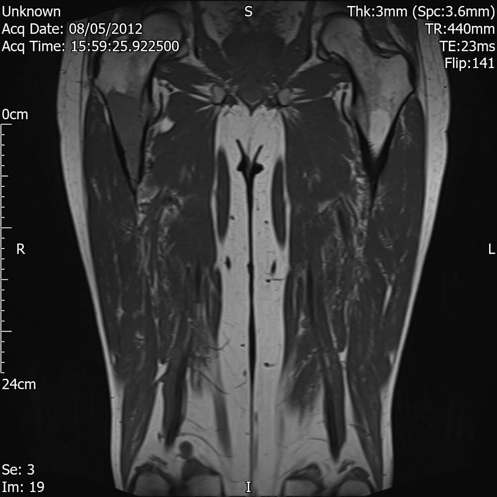
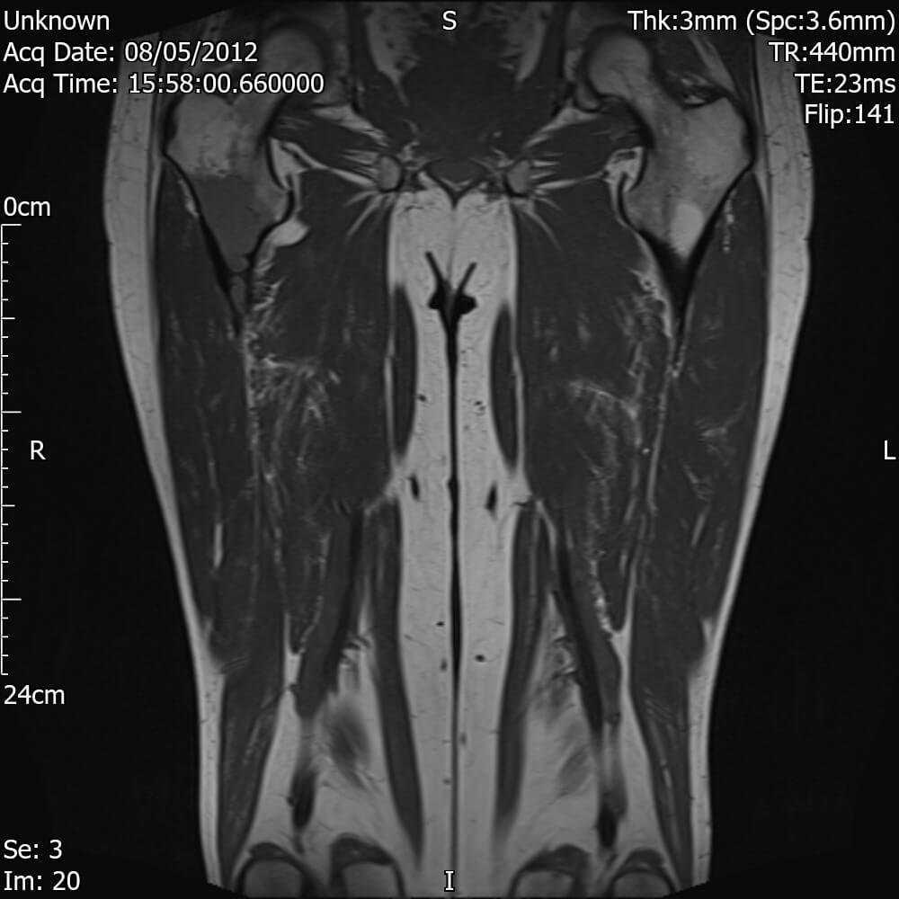
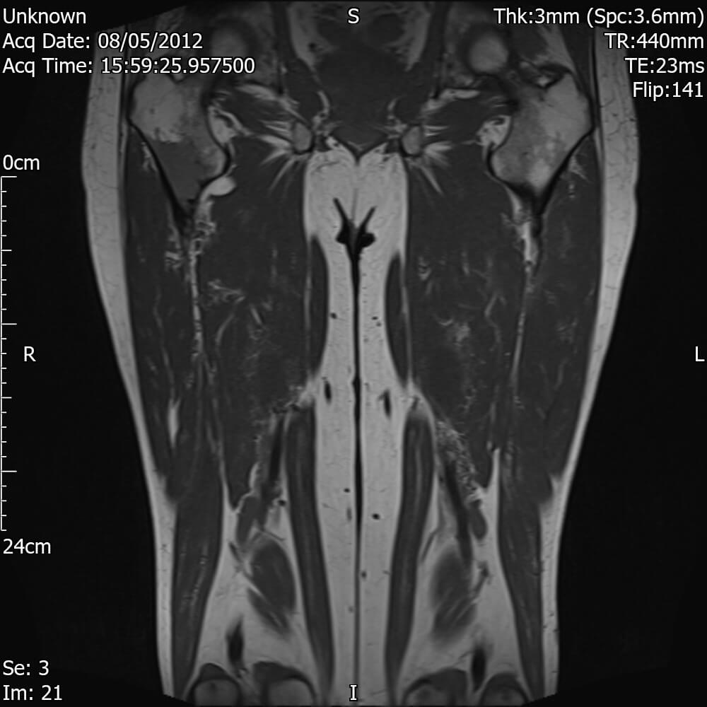
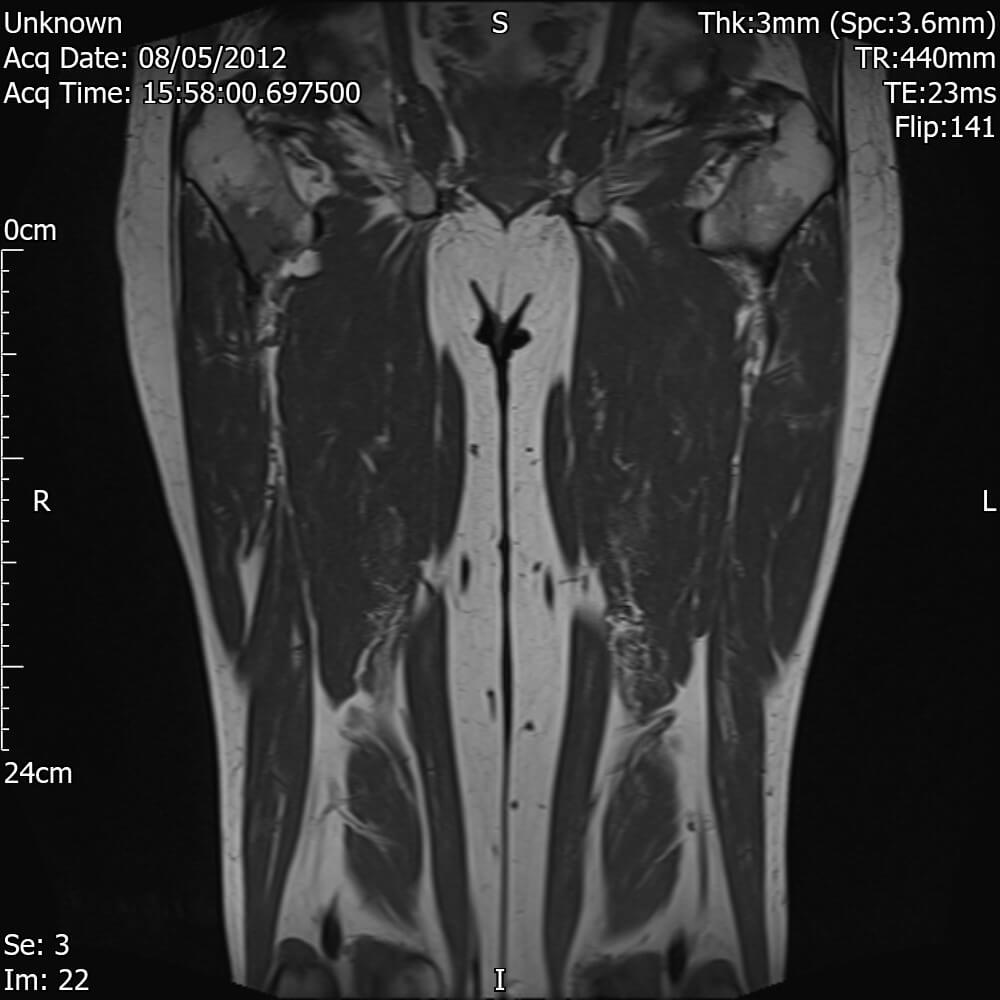
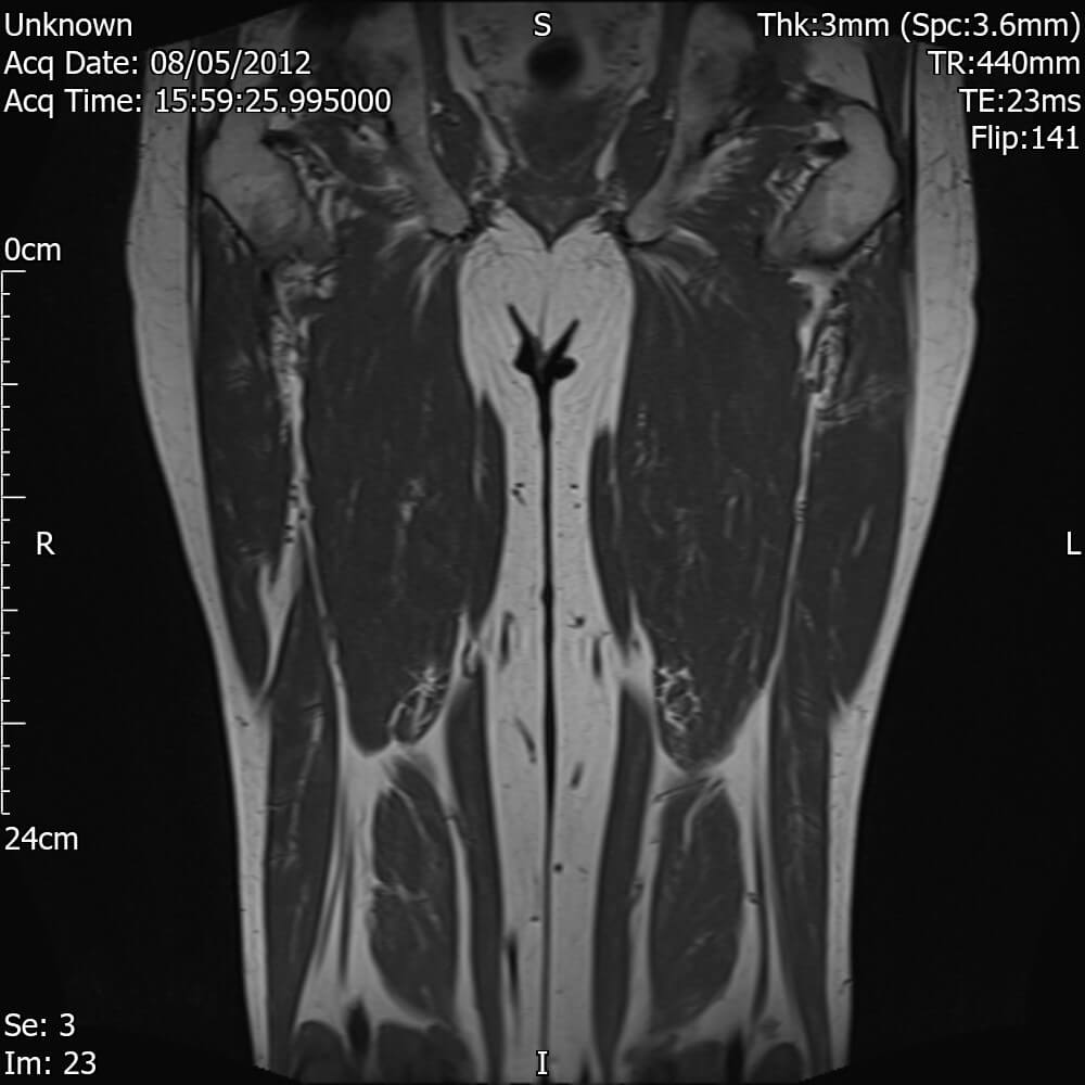
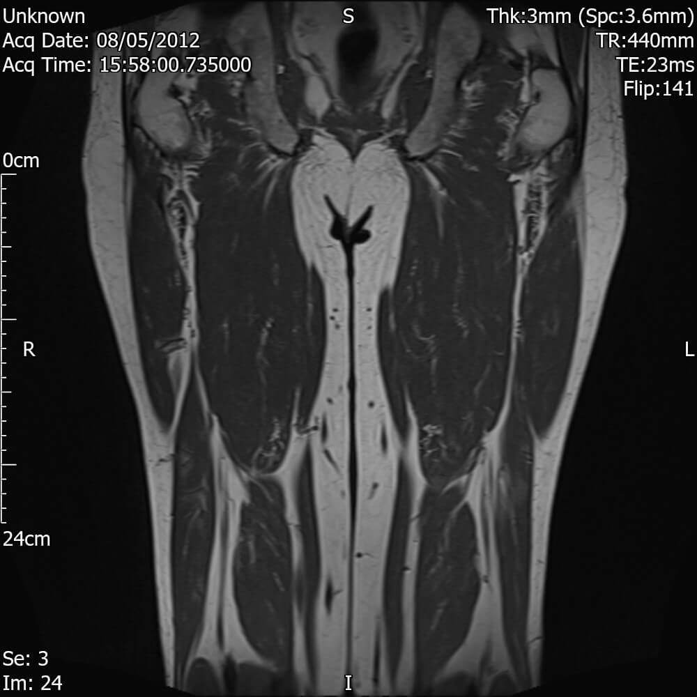
MRI Femur (Thigh) R
There is a medullary abnormality in the proximal right femur over a length of 11 cm which is intermediate in signal on T1/high signal T2 which causes thinning/erosion of the bone the cortex. Though there is a relatively sharp zone of transition the appearances are very suspicious of neoplasm. No marrow abnormality is identified in the distal right femur or in the left femur.
Suggest myeloma screen and bone biochemistry in the first instance. It is probably worth arranging CT of chest, abdomen and pelvis and discussing with orthopaedics whether CT-guided bone biopsy is the next most appropriate step.
Patient then undergoes a bone biopsy – Confirms a plasmacytoma / myeloma
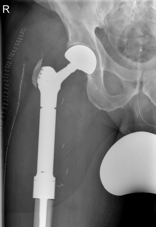
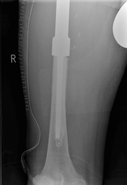
Surgical intervention
- Endoprosthesis replacing the right hip and proximal femur
- Patient then continues to have regular reviews under Haematology including CT scans, Chest Xrays and PET scans.
- 3 years after femur surgery, patient presents with xiphisternal pain.
- Patient is under Haematology, so undergoes a PET scan which shows a spinal compromise at T5. MRI is recommended.
MRI reports the following
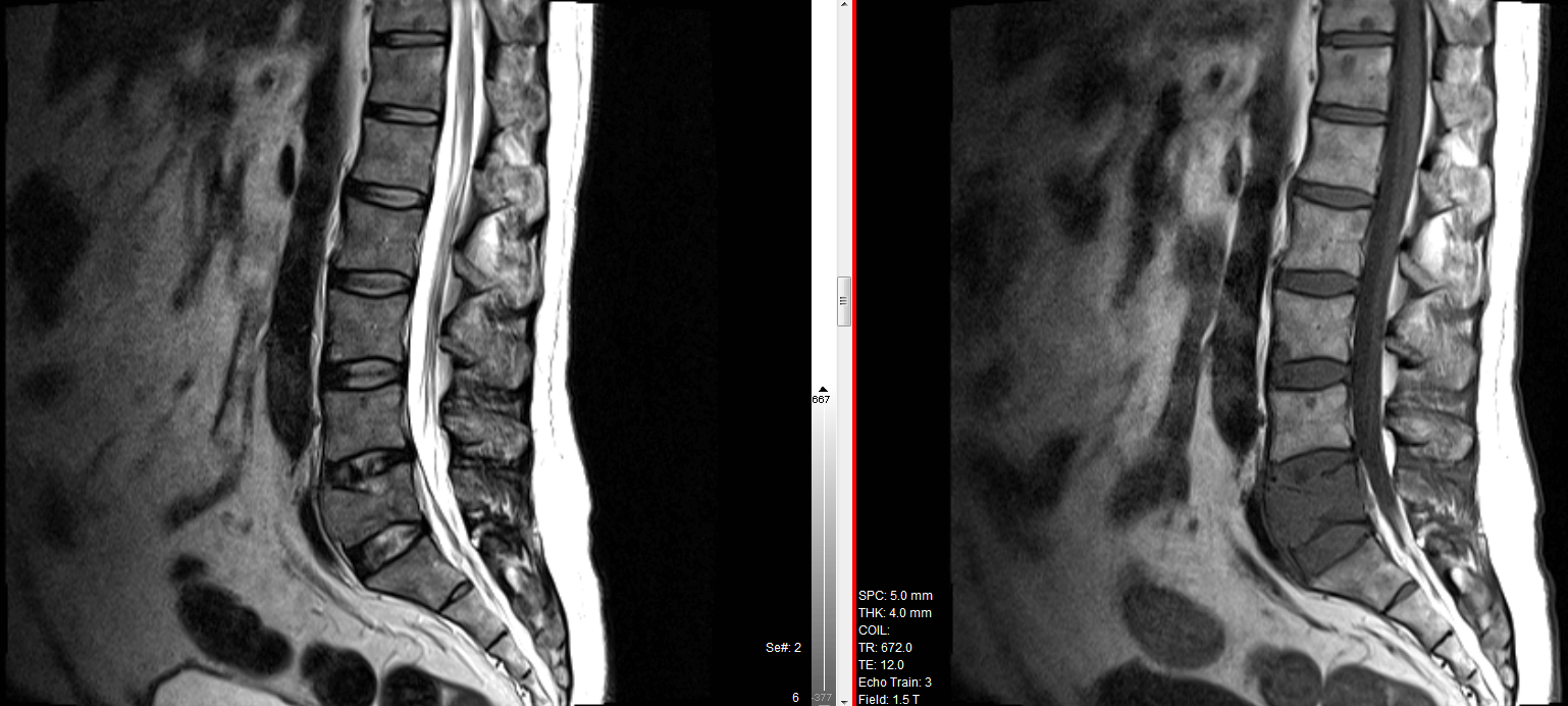
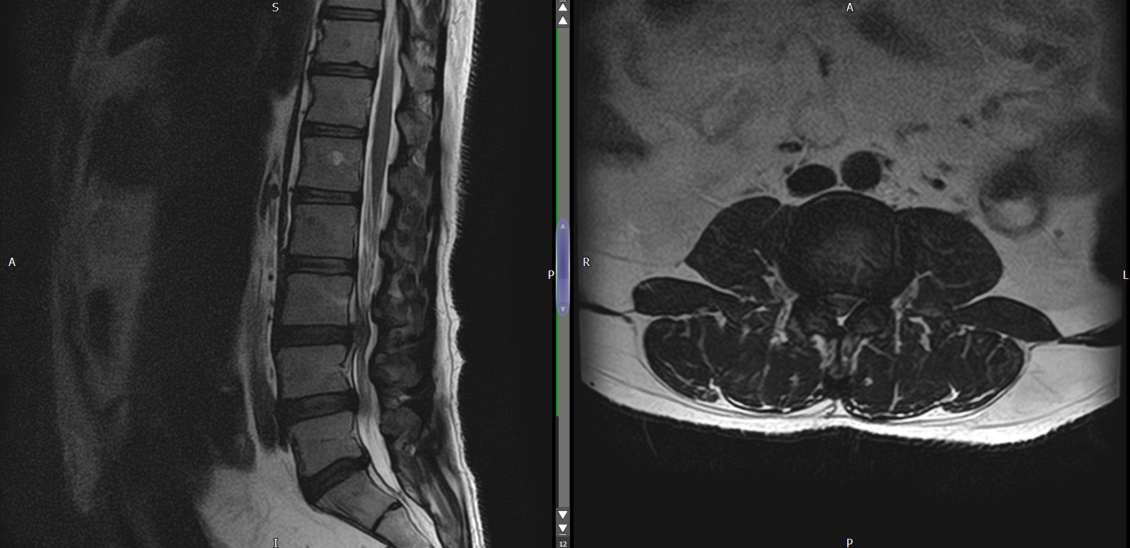
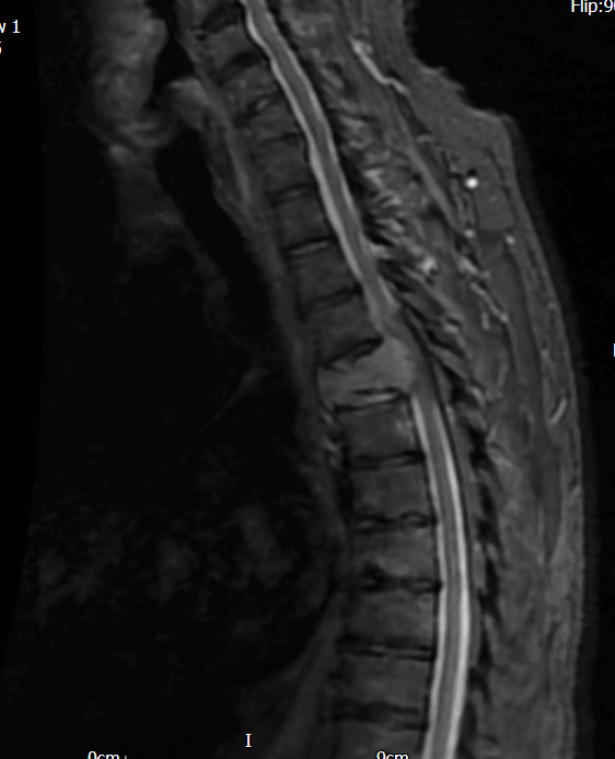
MRI Spine Whole :
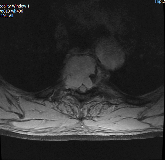
The bone marrow signal in T4 vertebral body is diffusely abnormal in keeping with tumour. There is loss of vertebral body height and tumour bulge both anteriorly and posteriorly. Posteriorly, tumour extends into the spinalcanal and compresses the cord. Tumour also extends into the spinal canal posterior to the lower half of T3 and the upper half of T5. There is also tumour involvement of adjacent right sided ribs and a tiny presumed focus of tumour anteriorly in T2 vertebral body.
CONCLUSION: Cord compression at T4.
Diagnosis – Multiple Myeloma with T4 osteolysis.
MRI images T4 level
Patient undergoes the following procedure.
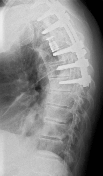
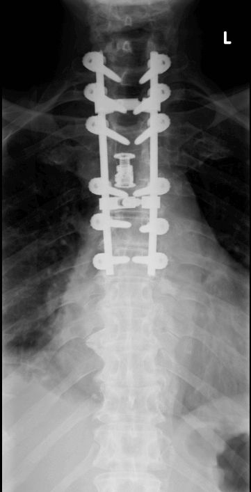
- Posterior instrumentation T1 – T7
- Costo transversectomy T4
- Subtotal corpectomy T4
- Fortify cage T3 – T5
- Reduction of deformity