Mass in Medial Clavicle | Cases
Published on Jan 15, 2021
Lump over Medial clavicular region close to SCJ
57 female Reports slow growing lump in chest region for over 5 years. No trauma. Gets some shoulder pain. Has been having osteopathy for shoulder.
Full shoulder ROM on examination. Non tender lump. Mild SCJ tenderness. Clinician requested MRI scans.
Retrospective and current investigations below.
X-ray April 2017
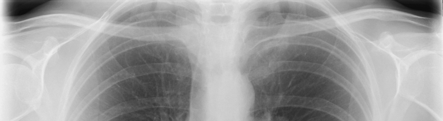
X-ray Feb 2020
Report: There is expansion and sclerosis of the medial third of the clavicle raising the possibility of sclerotic metastasis from for example, carcinoma of breast. Further imaging advised, initially chest x-ray and specific views for right clavicle.
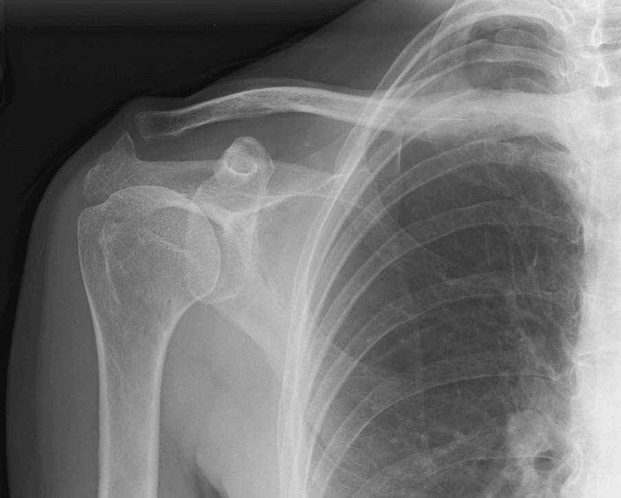
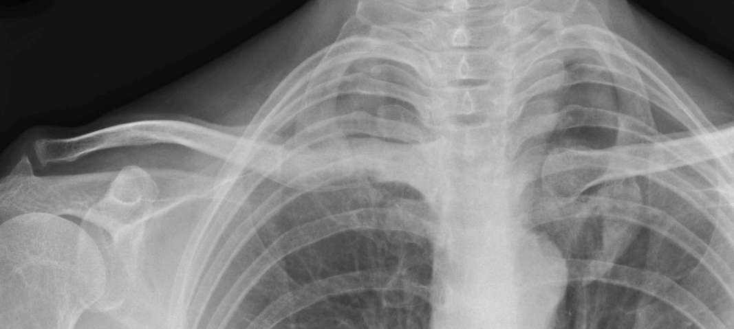
X-ray Aug 2020
X-ray Jan 2021
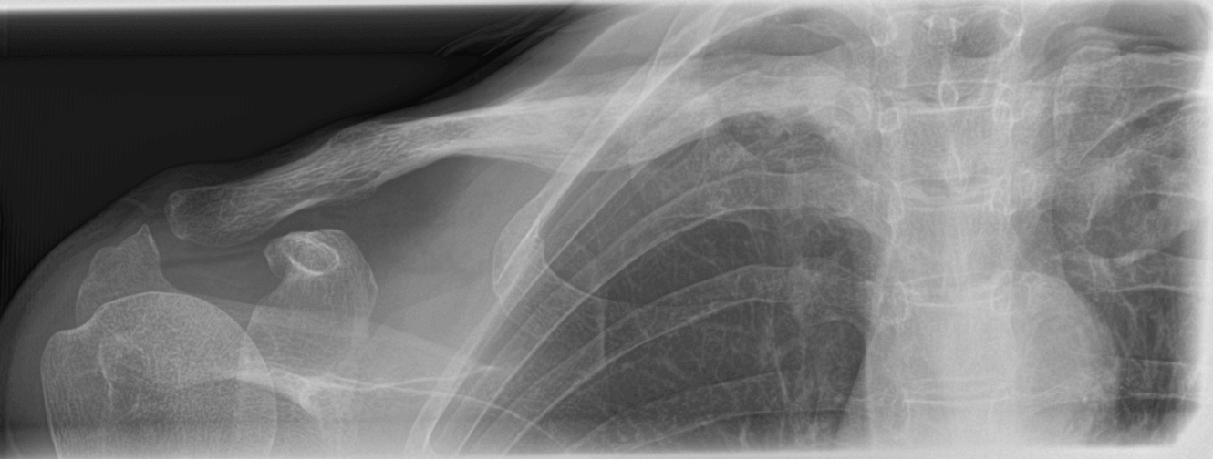
Report: There has been some slightly progressive expansion of the medial and middle thirds of the right clavicle when compared with the prior x-ray dated 1 February 2020 and there is also a new/progressive soft tissue swelling at the level of the middle third of the clavicle. The previously demonstrated sclerosis within the medial third of the clavicle has decreased and the bone is slightly more lucent on the current examination.
There is no other bony abnormality within the field of view. No acute fracture is identified. I also understand there is no history of prior trauma to suggest a healing fracture.
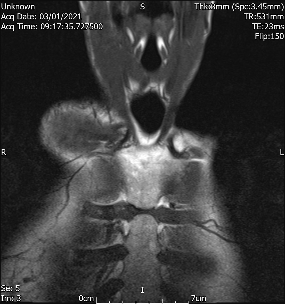
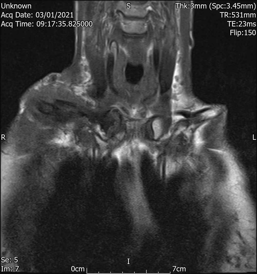
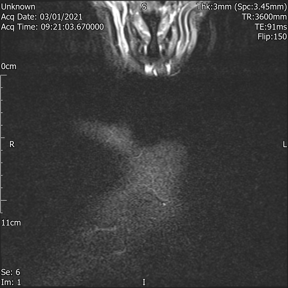
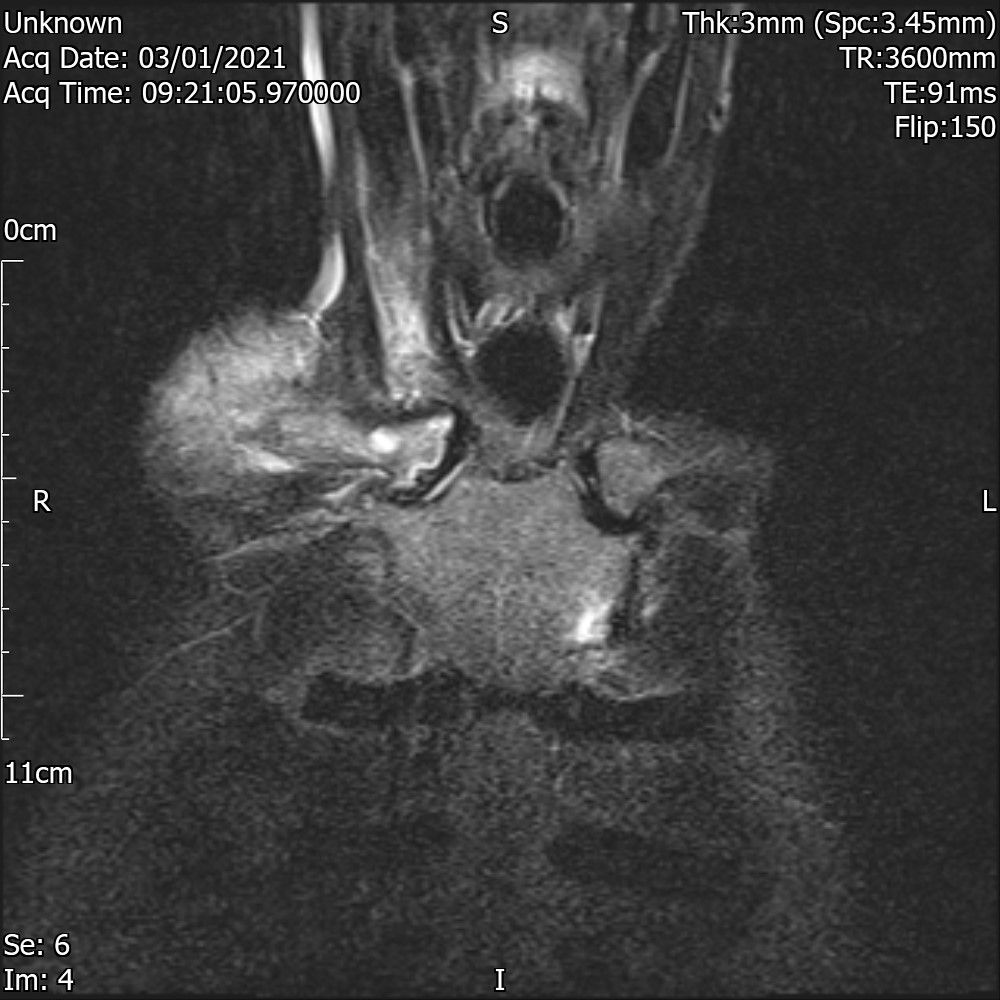
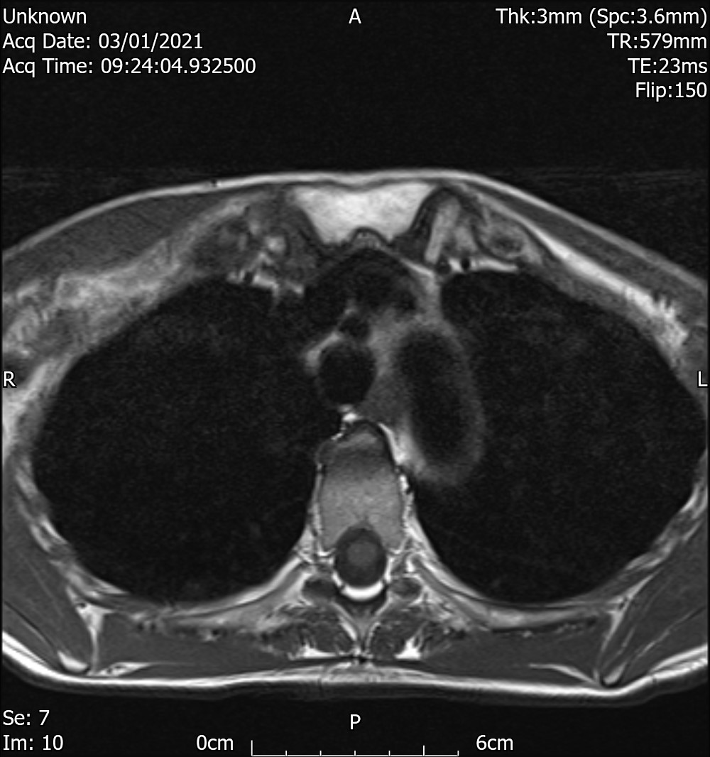
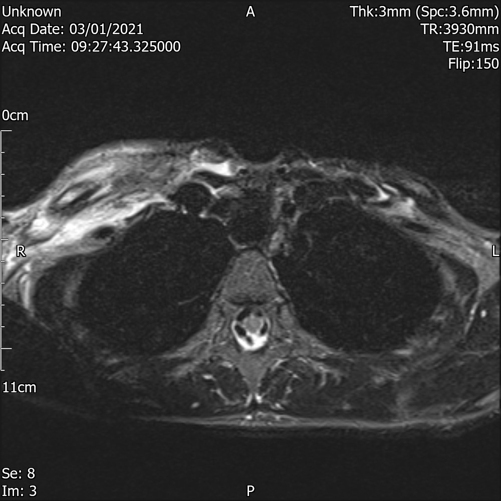
MRI T1
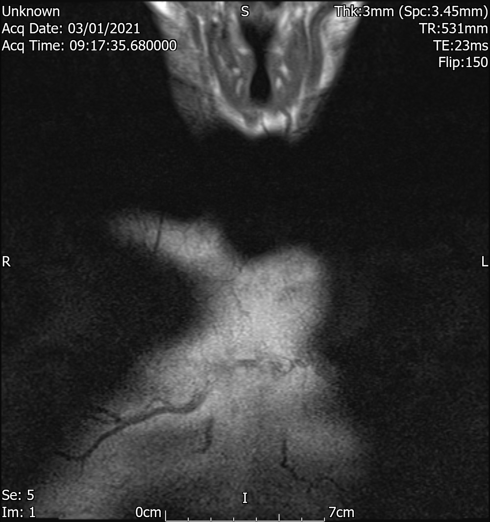
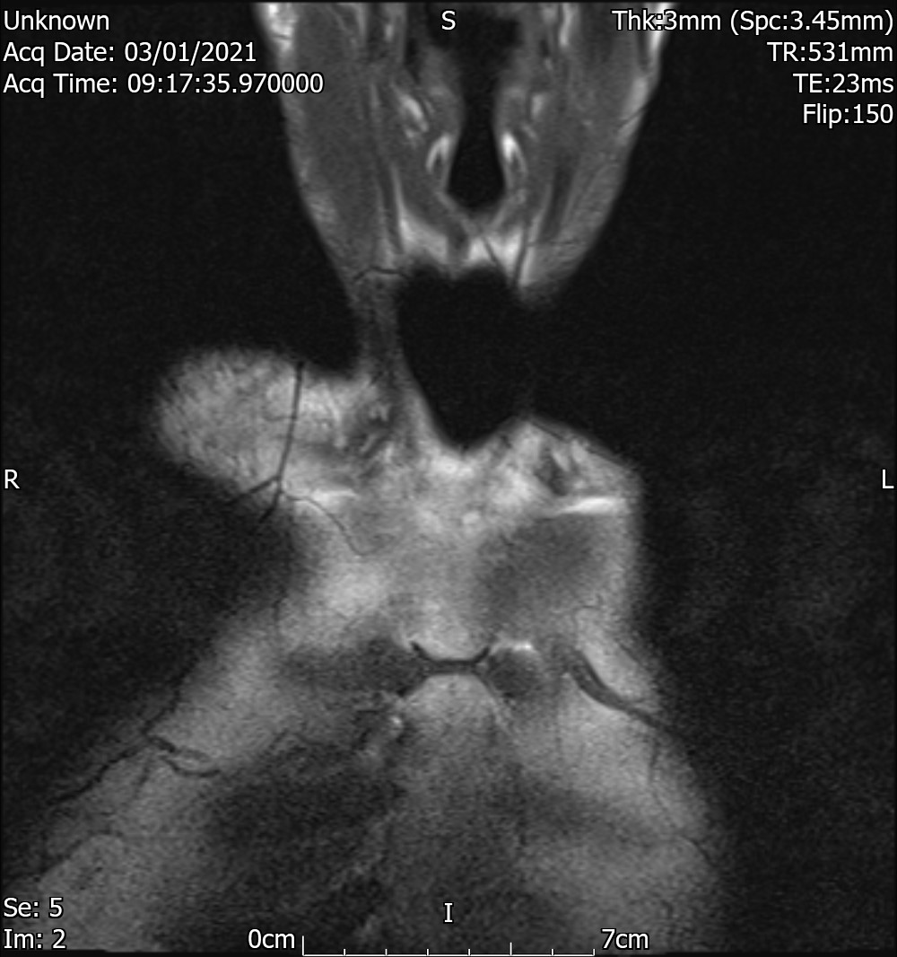
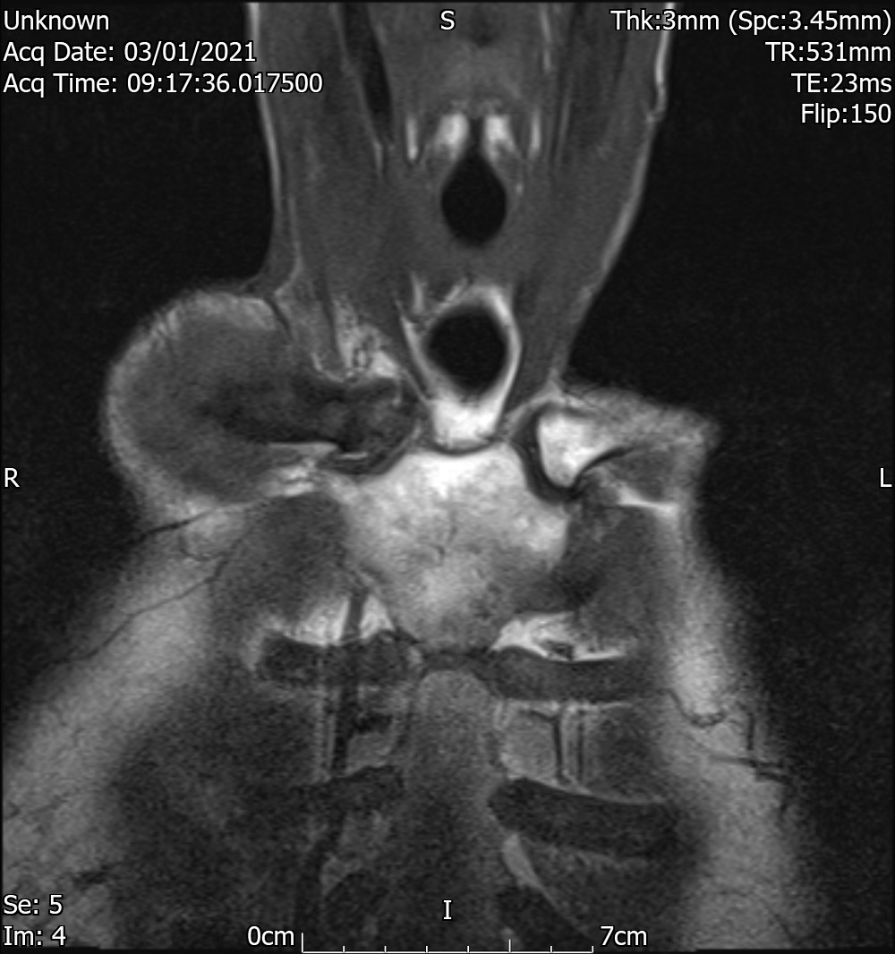
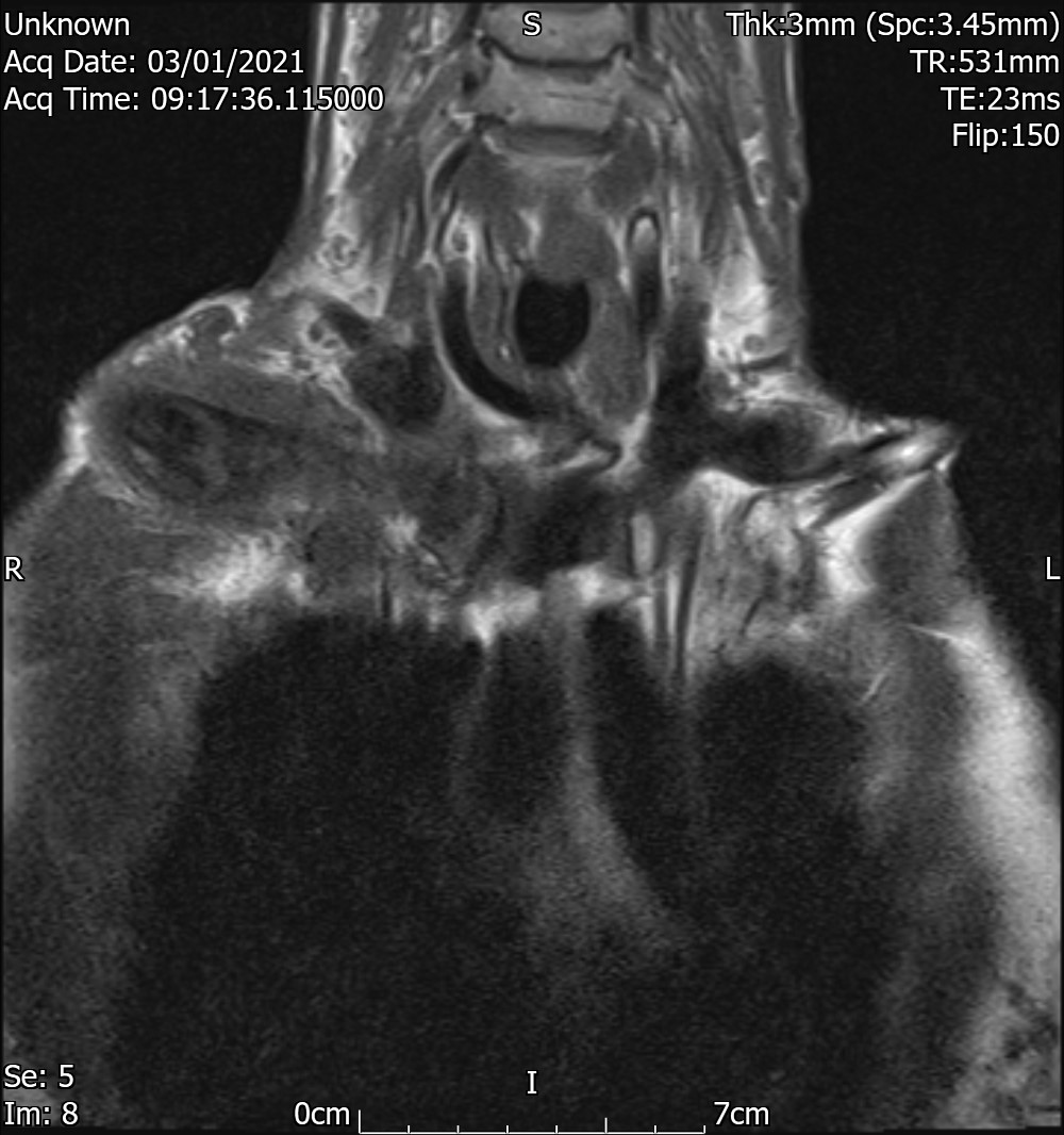
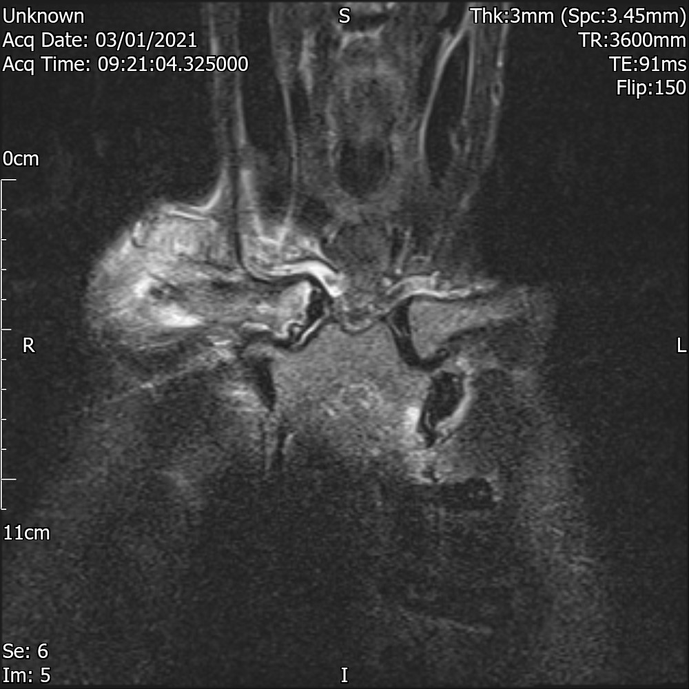
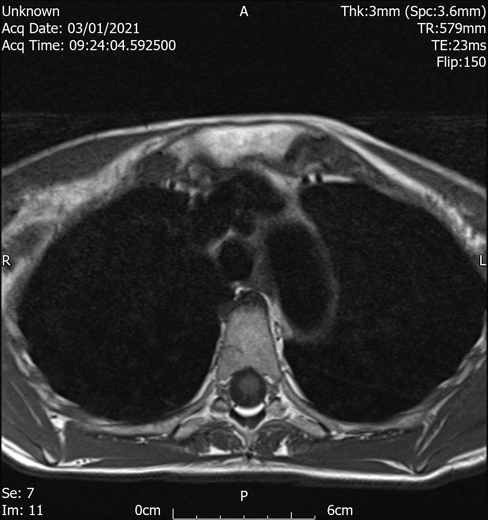
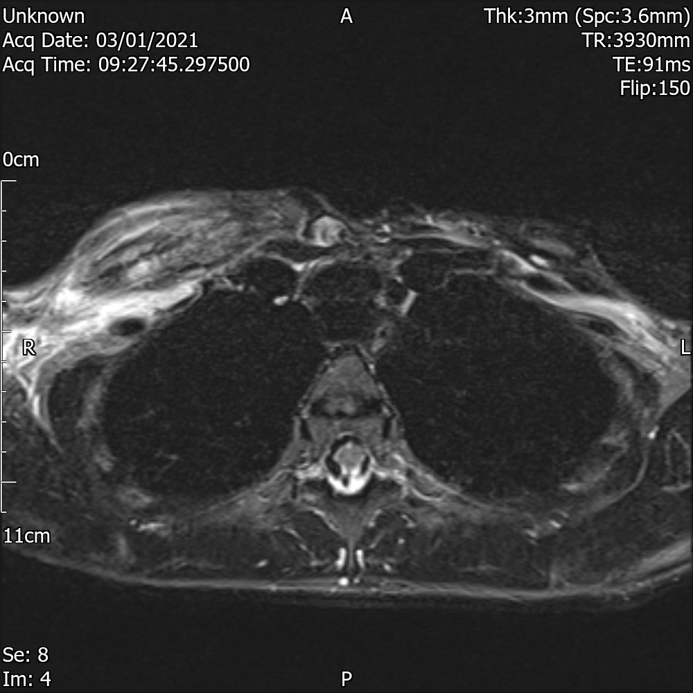
MRI T2
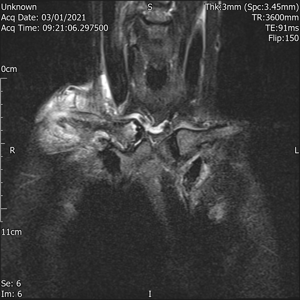
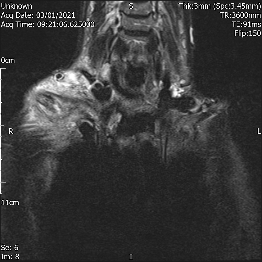
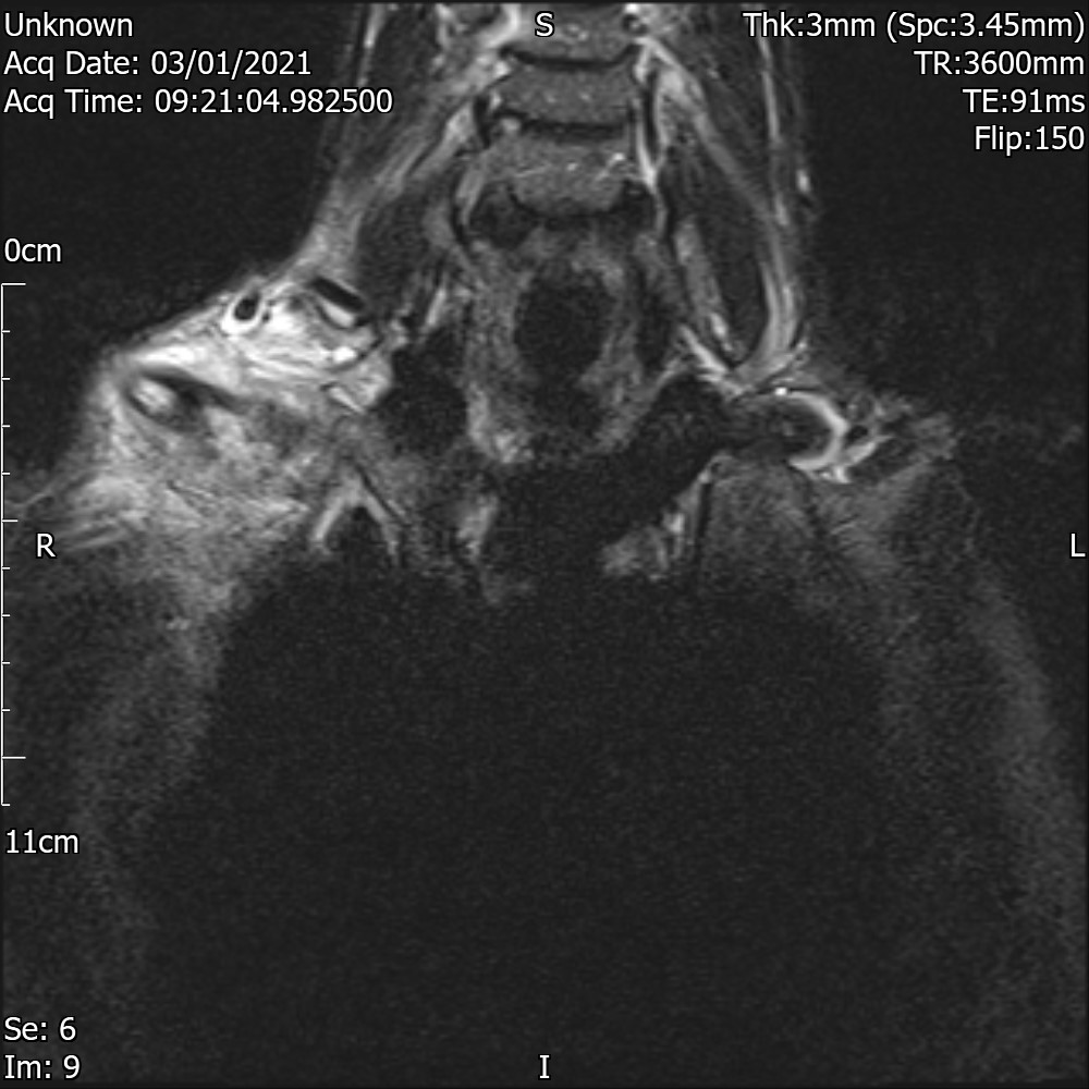
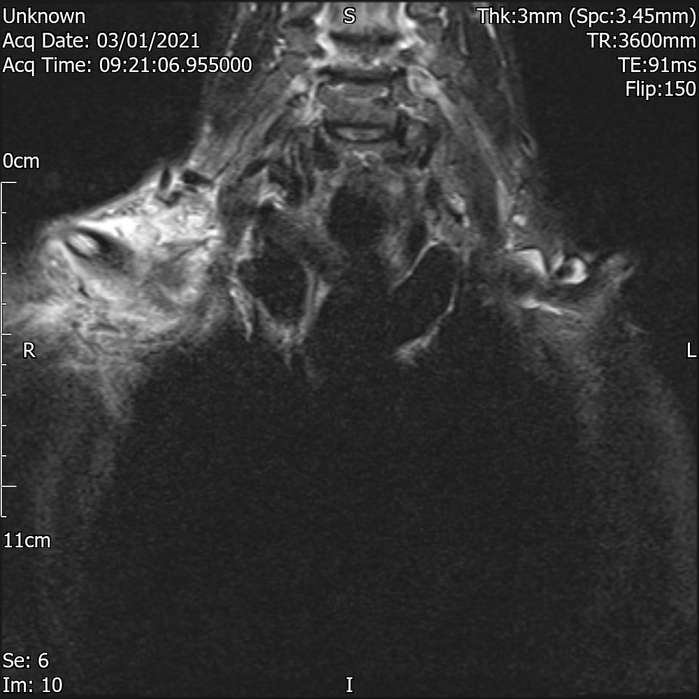
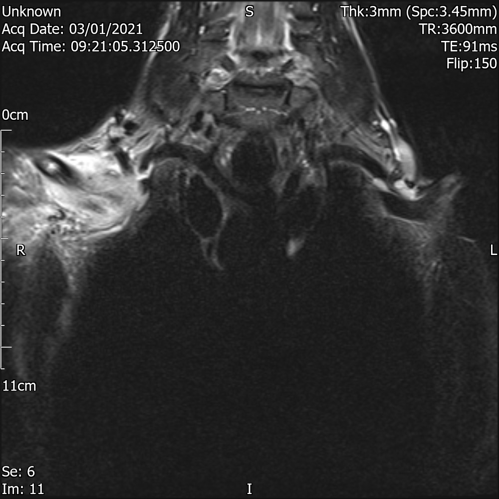
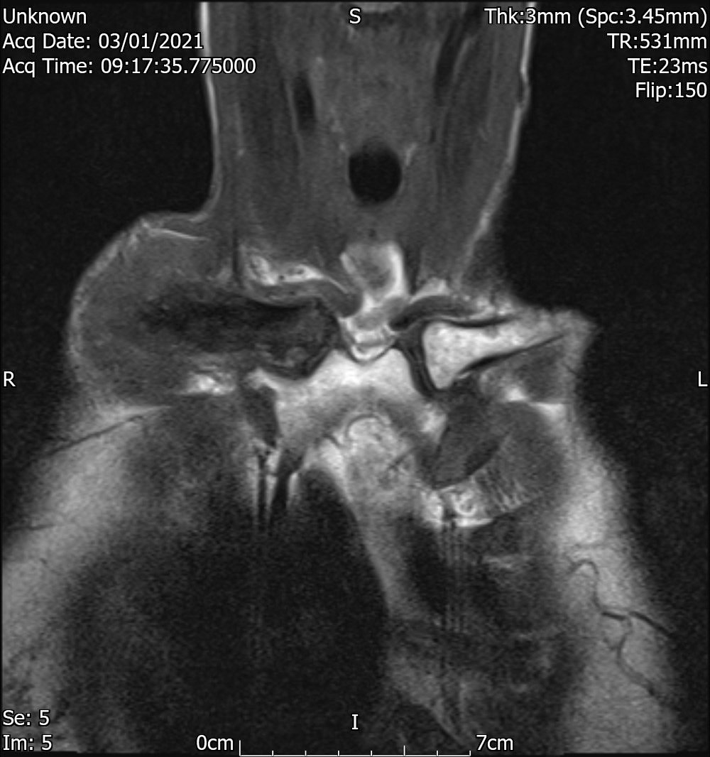
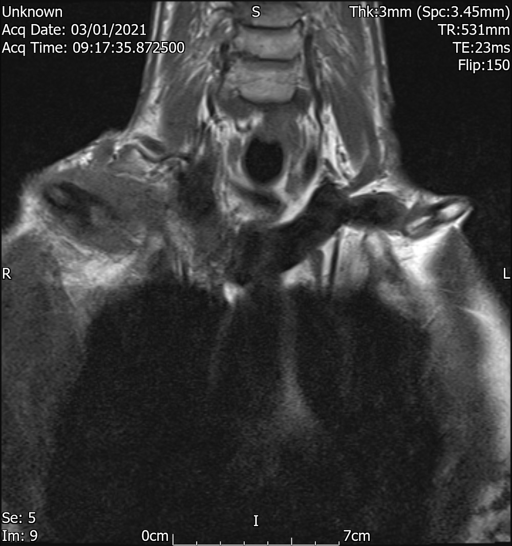
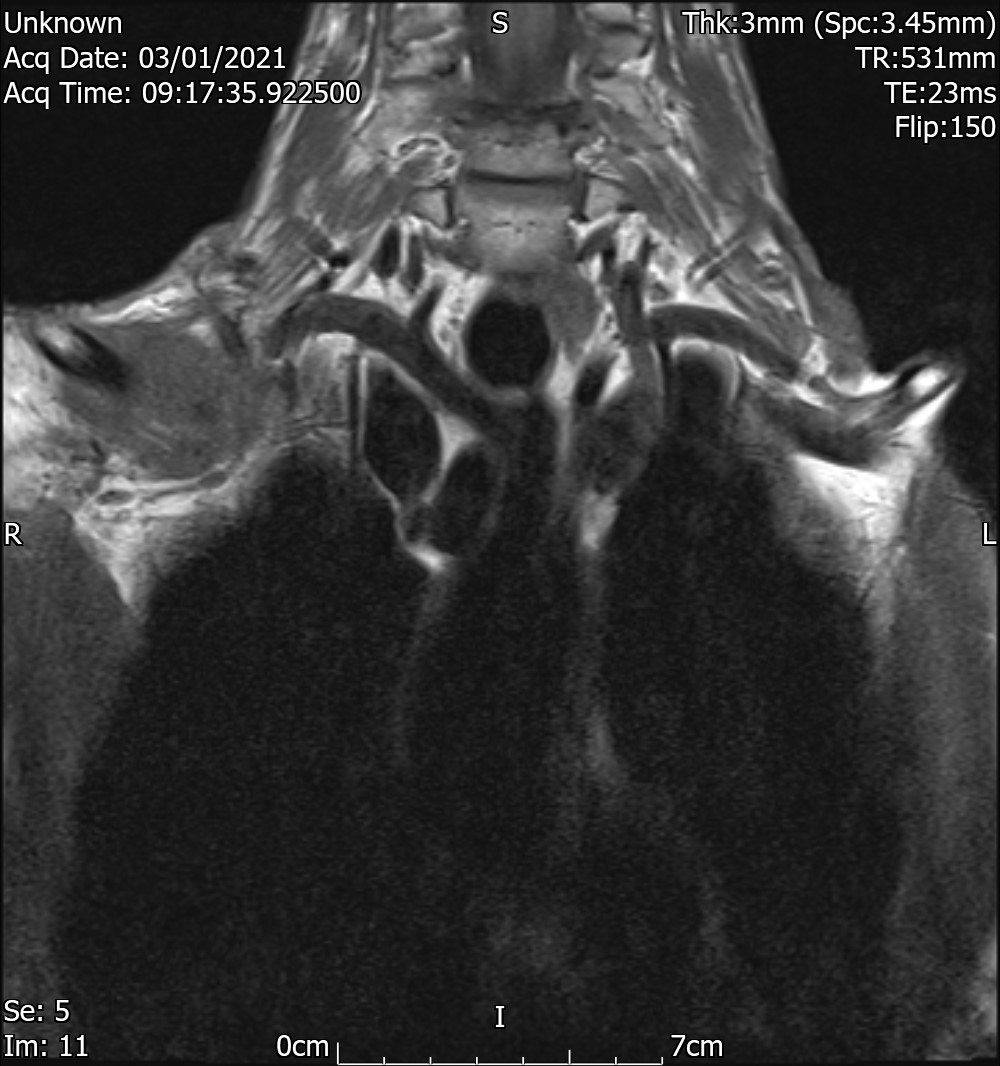
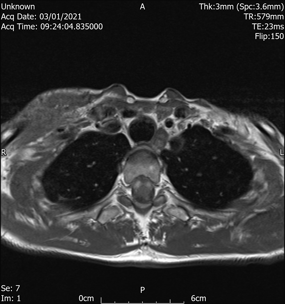
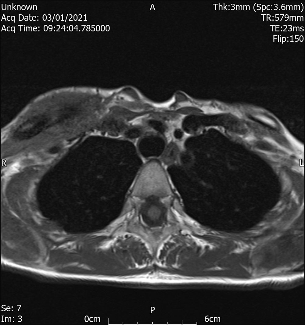
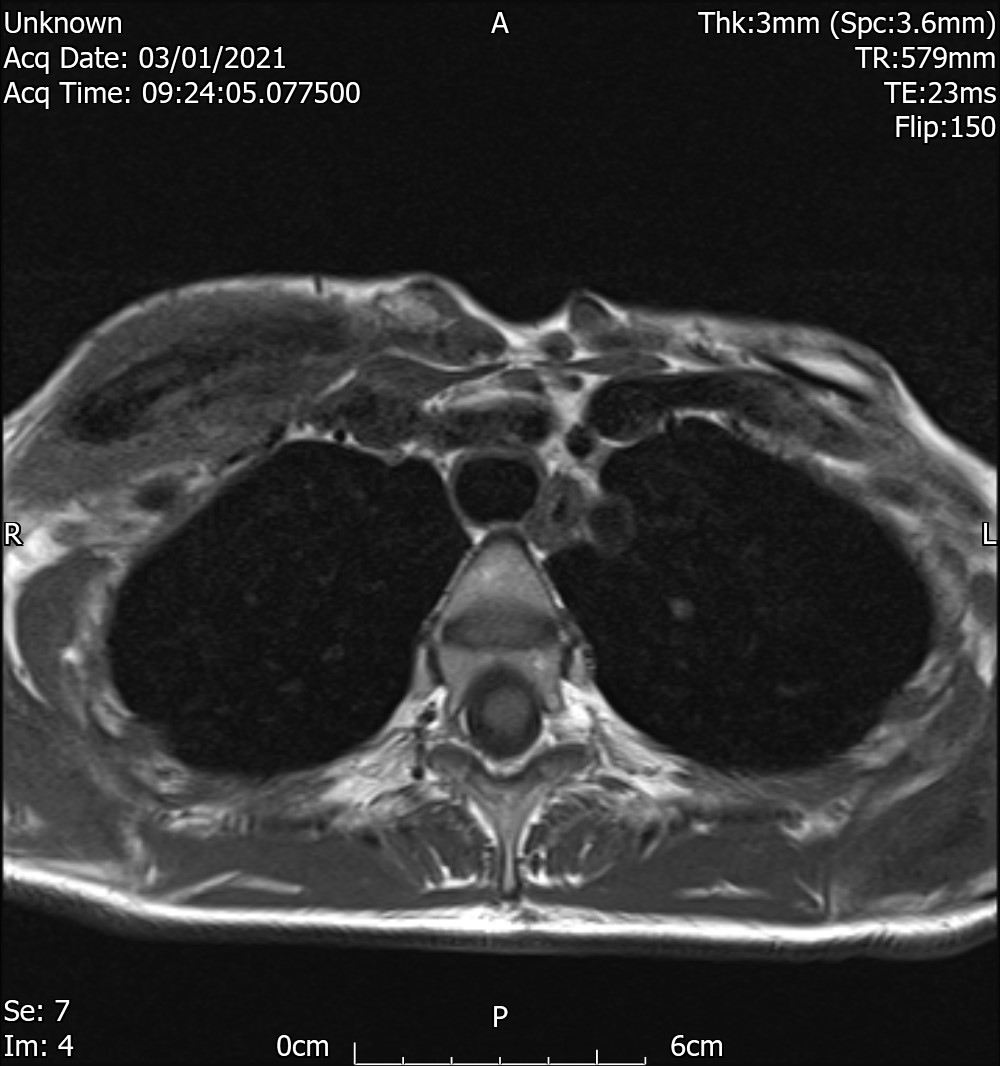
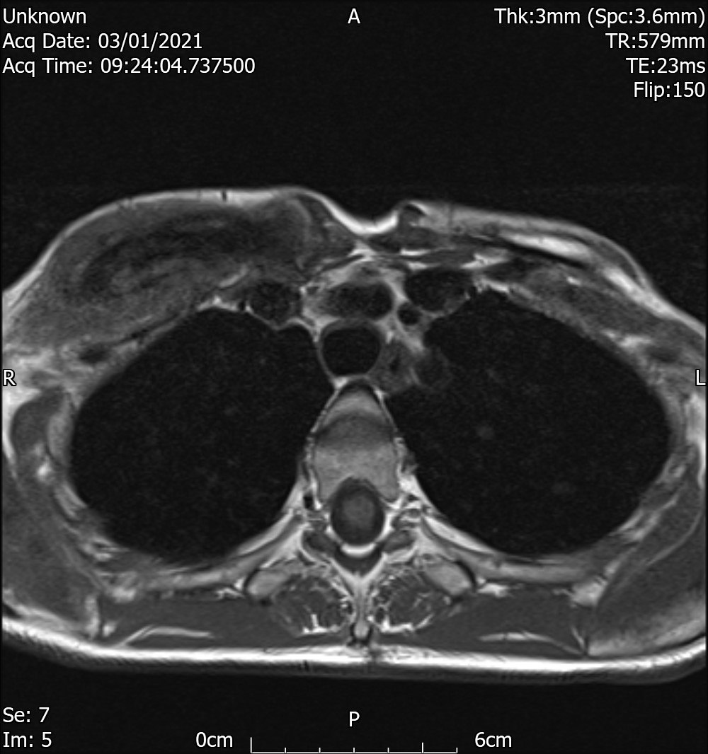
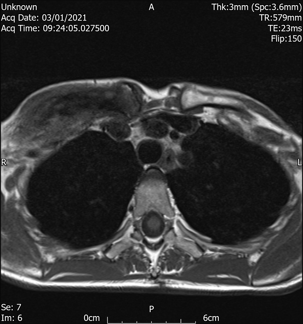
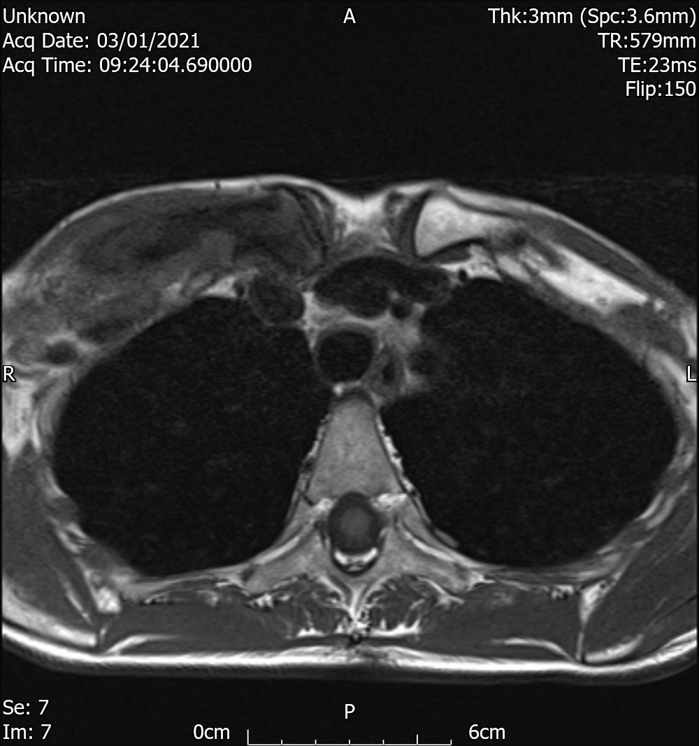
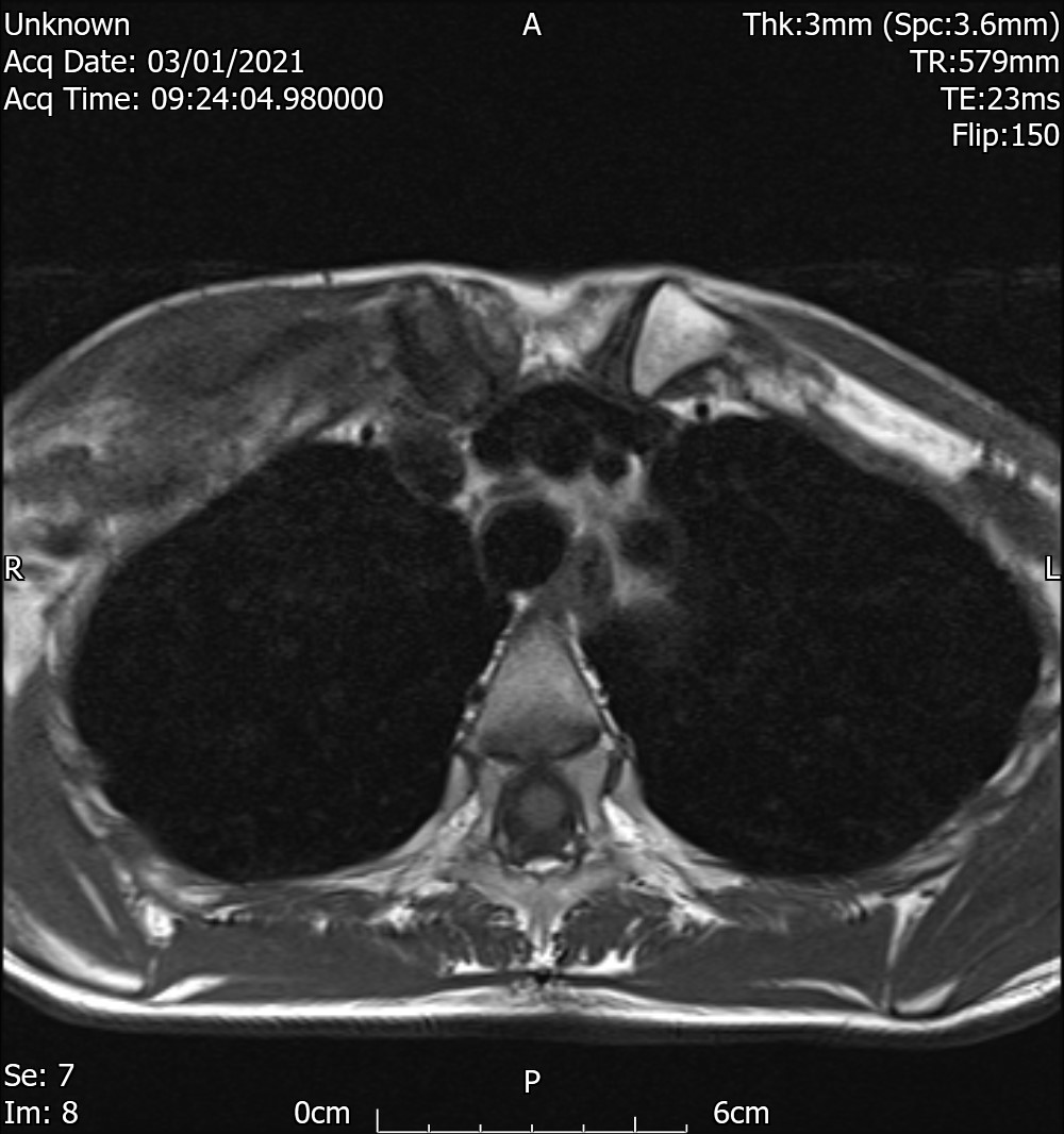
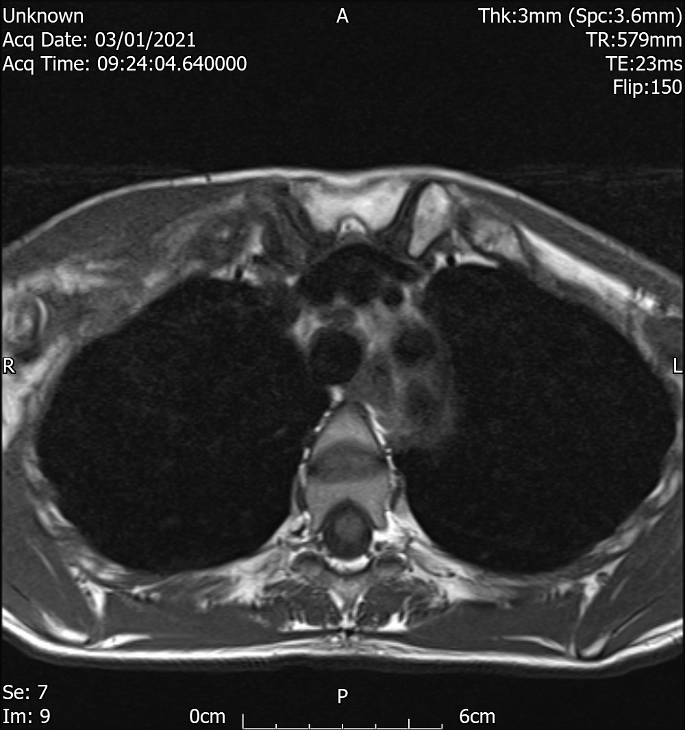
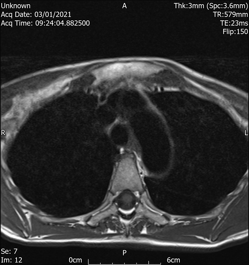
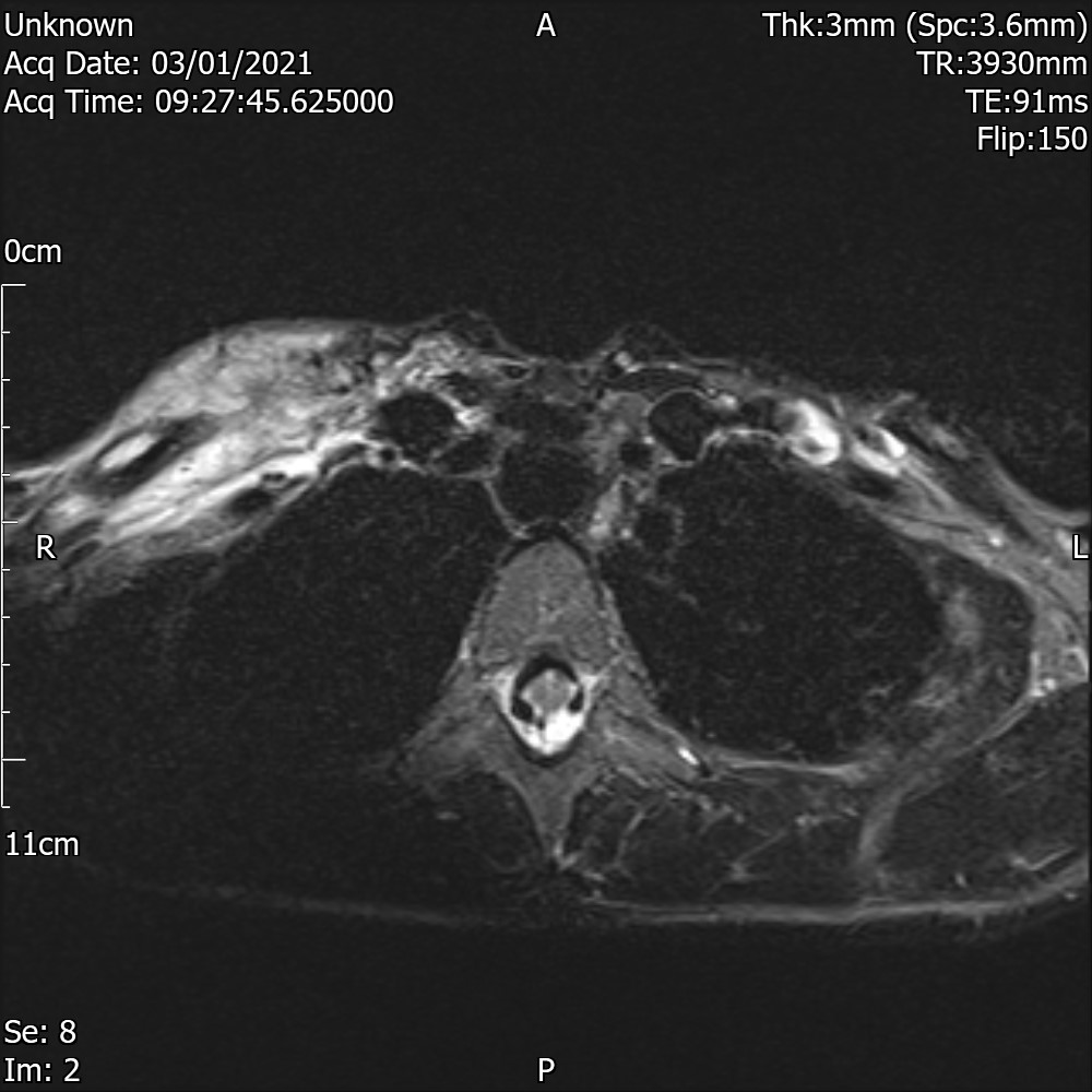
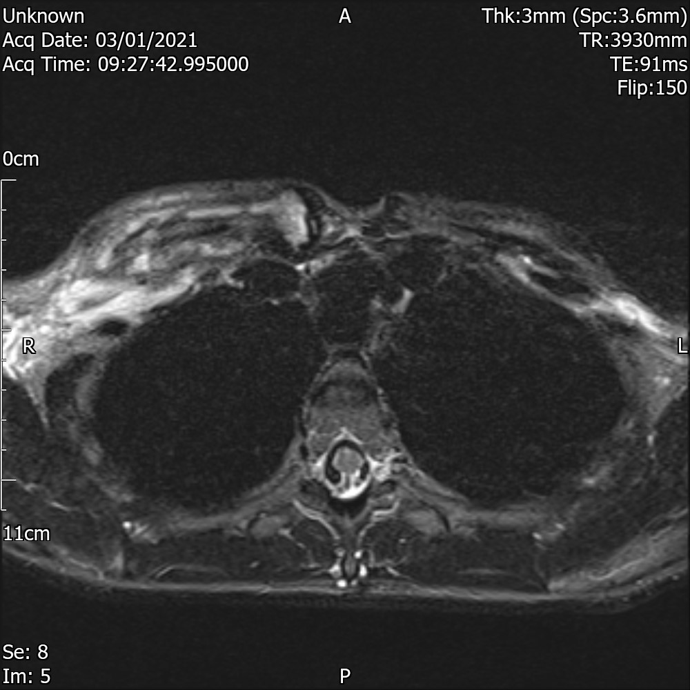
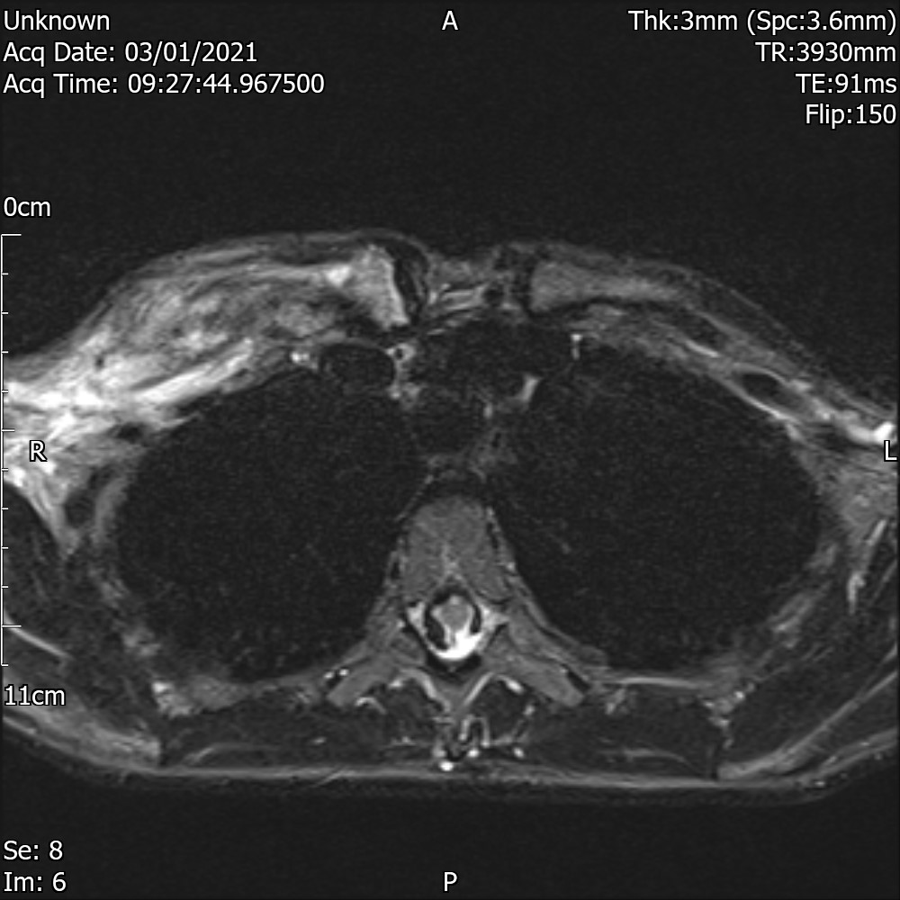
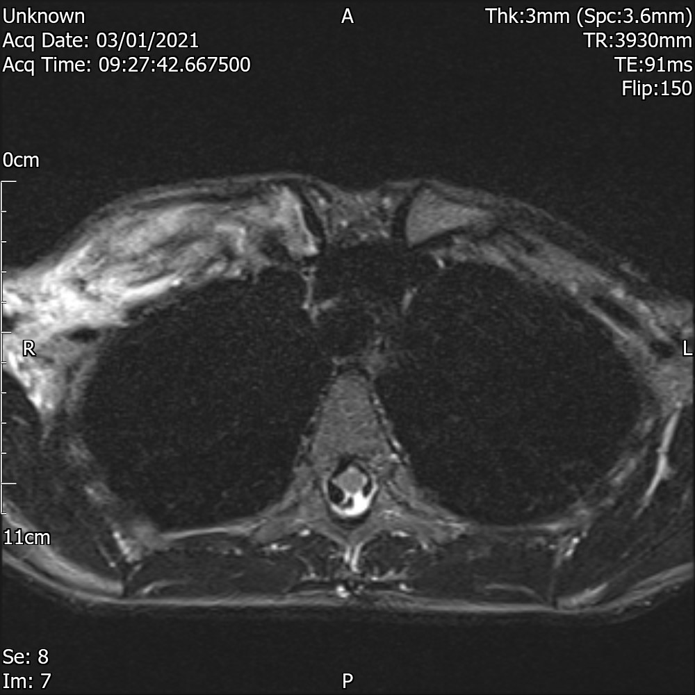
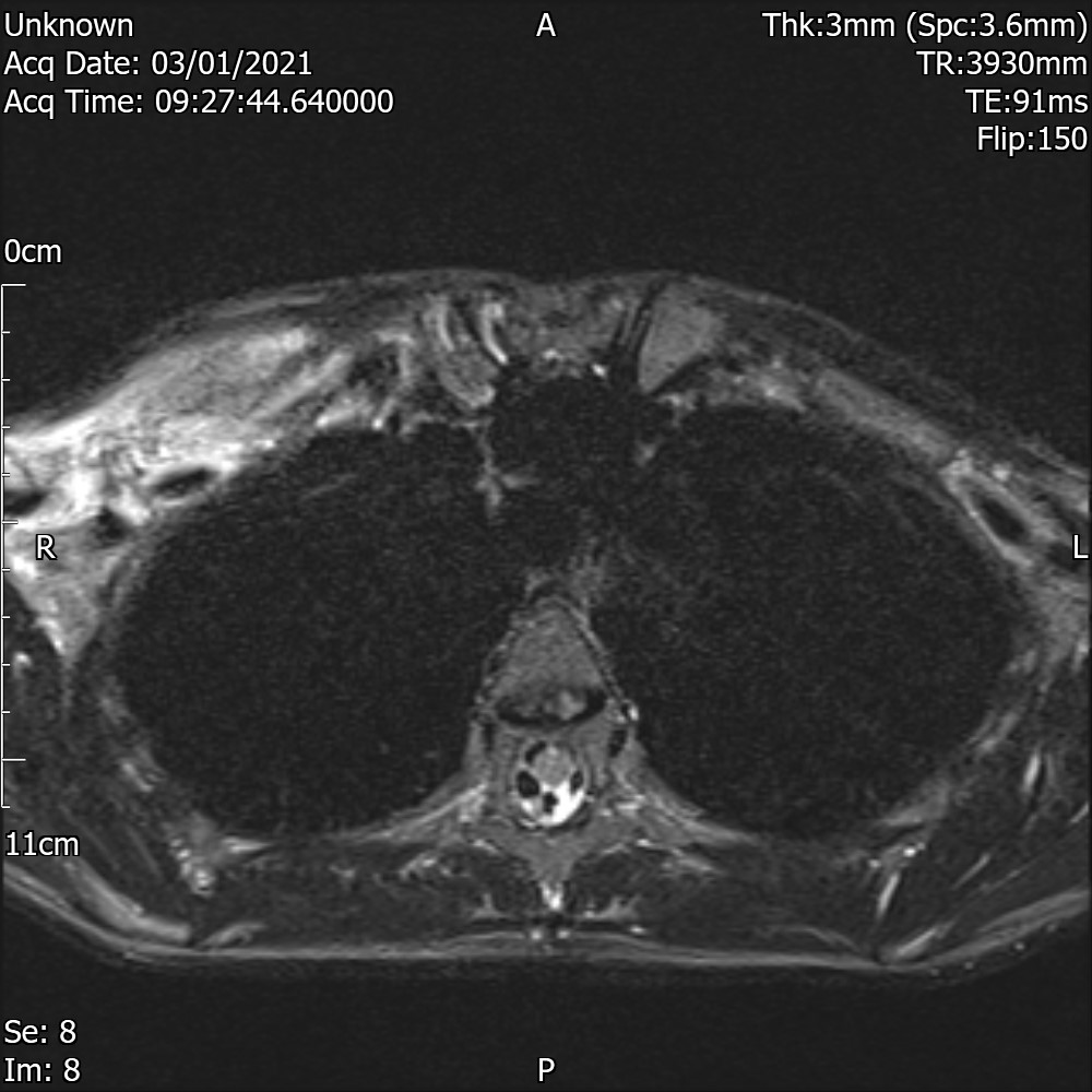
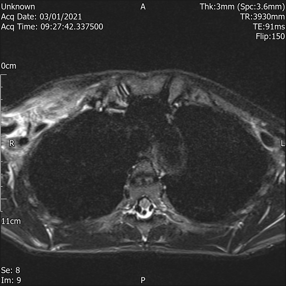
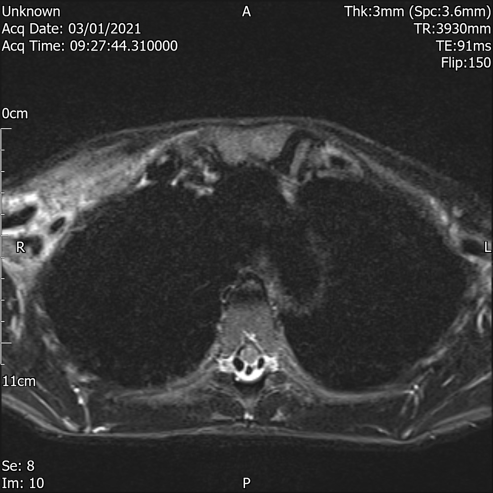
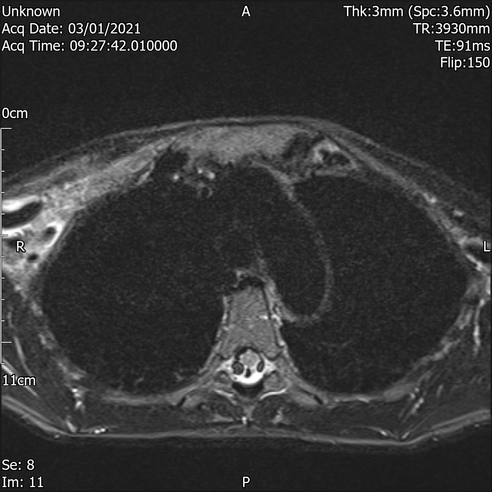
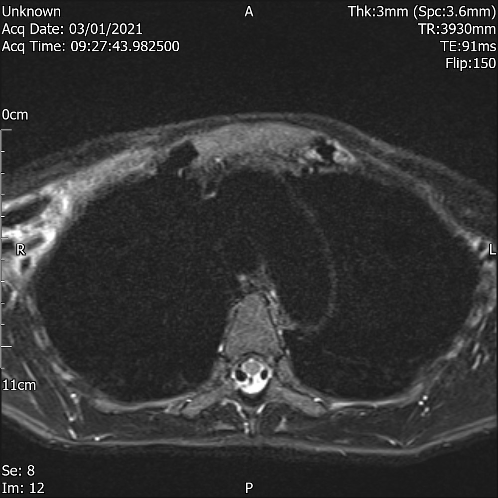
MRI T1 Axial

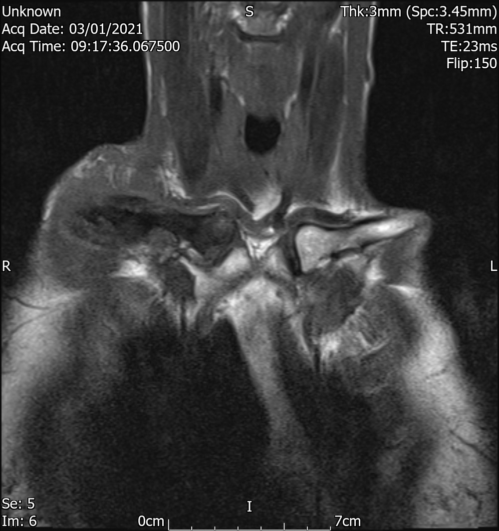
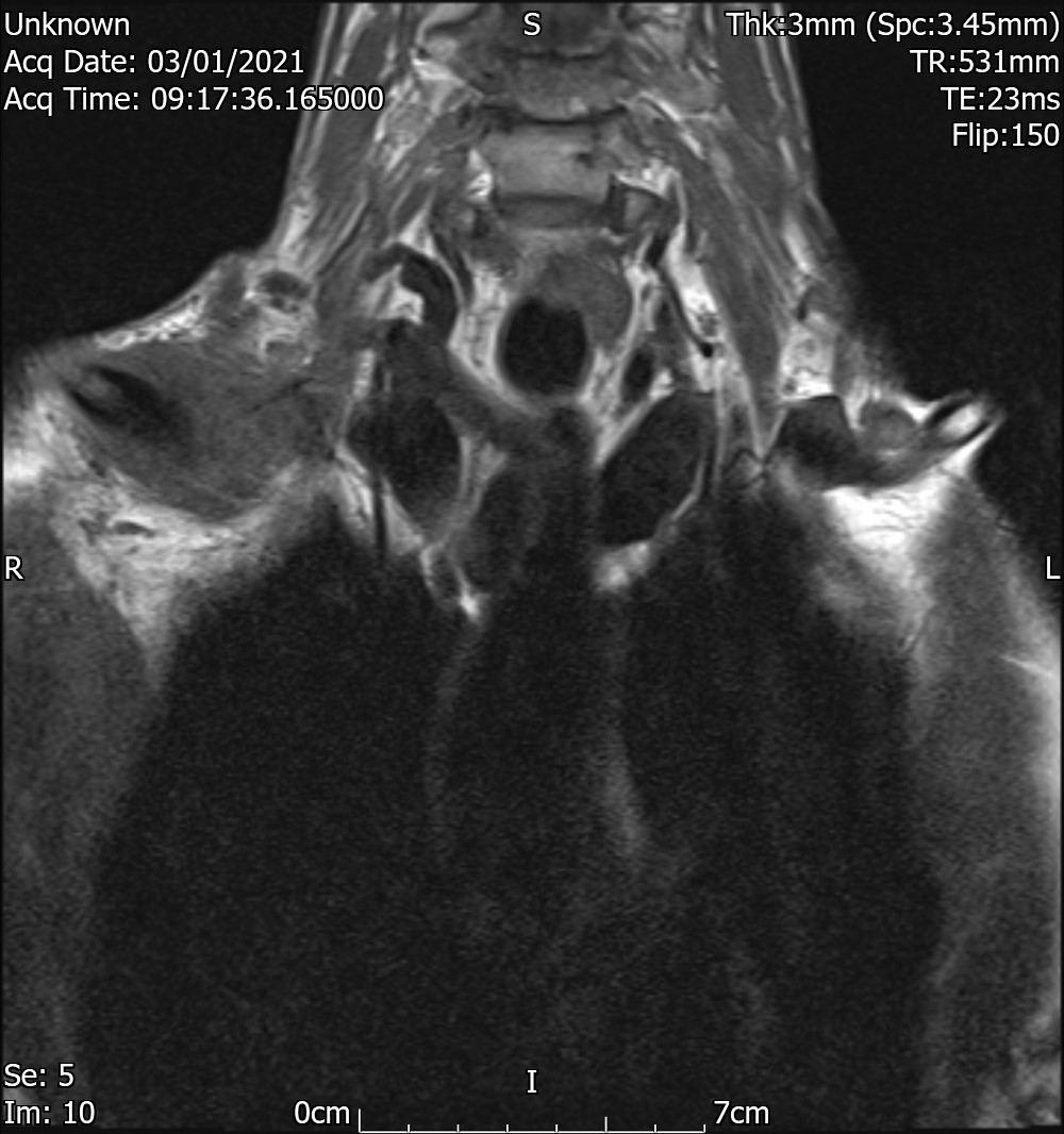
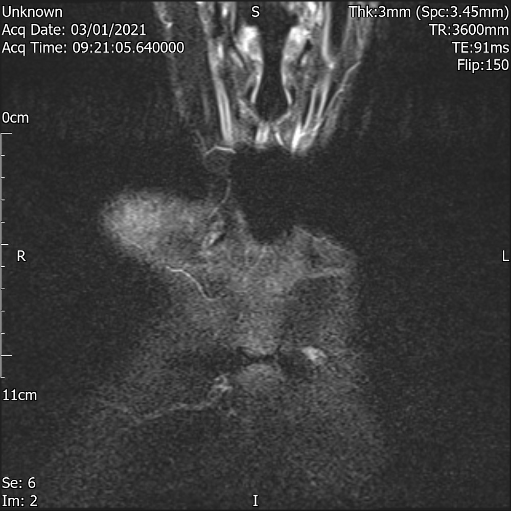
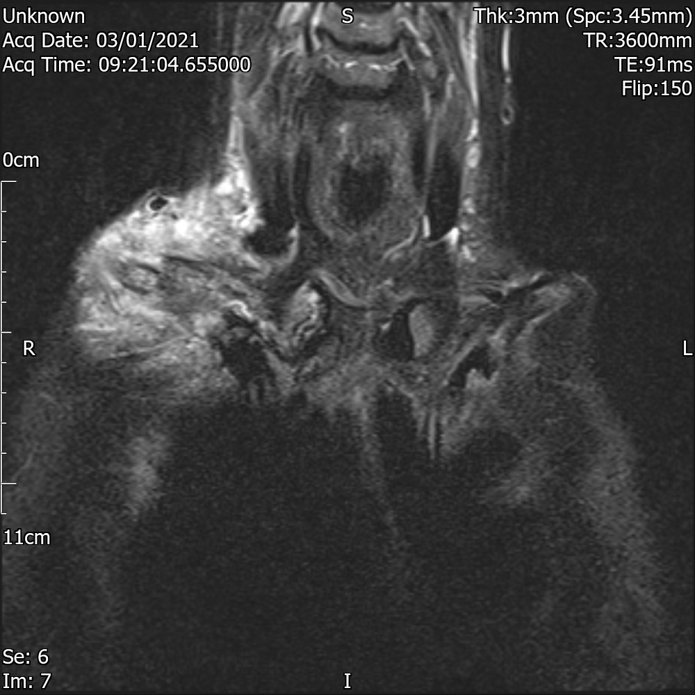
MRI Trim Axial
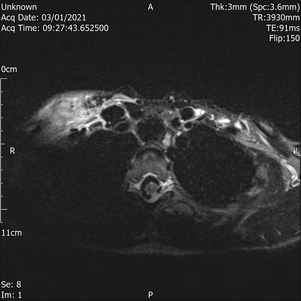
MRI Report
No current plain films for comparison. Most recent examination 21 February 20 is abnormal.
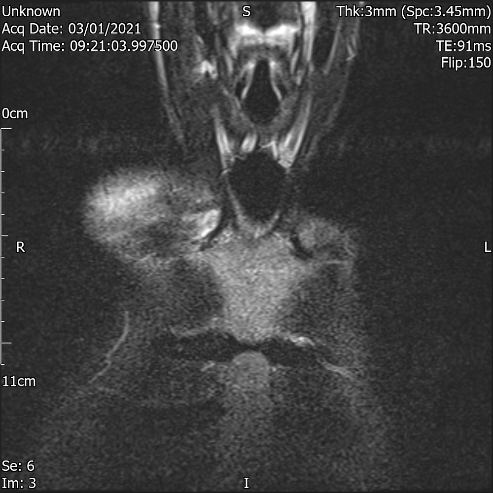
The medial third of the clavicle is expanded and ill-defined with loss of the cortical medullary definition. My first thought would been healing fracture but the possibility of bone tumour or the possibility of Paget or Paget sarcoma would a consideration. Rule one is always that crucial that any MR assessment of a bone lesion is associated with contemporaneous plain film.
The MR shows very abnormal medial half of the right clavicle. There is mild oedema through the through the visualised bones. There is a lesser lamina change around the bone with circumferential soft tissue swelling although discrete destructive mass with ample is not demonstrated. There is a little oedema within the sternoclavicular joint. There is no obvious changes elsewhere and involvement the clavicle in the absence of other findings makes this unlikely to be part of a SAPHO syndrome.
Concealed fracture is a consideration but I understand there is no provided history of trauma and clavicular fractures tend to be distal rather medial third.
There is too much soft tissue swelling for Pagets which is also an increasingly uncommon
finding. Soft tissue changes are also not typical for tumour. Parosteal soft tissue tumour mass is possible. The coronal images look very suspicious for fracture however a tumour must remain a concern.
Repeat plain film and CT assessment is recommended which may allow for further interpretation of the MRI scan.
CONCLUSION
Abnormal clavicle with suspicion that this is fractured on the coronal imaging. Complex bone and soft tissue changes are noted but not definite for tumour although the appearances are not consistent with infection. Repeat plain film and imaging and CT assessment is required.